AMD Pictures and Videos: What Does Macular Degeneration Look Like?
What Are Fundus Photographs?
Ophthalmologists can only see the effects of age-related macular degeneration (AMD) by looking closely at the retina. They use a special camera to shine light onto the back of the eye (fundus) and take pictures. Ophthalmologists call these pictures fundus photographs.
Dry AMD Fundus Photographs
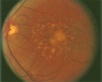
Drusen are fatty deposits under the retina. Hard drusen are not a sign of AMD. Soft drusen are closer together and form larger deposits. They can be an early sign of AMD.
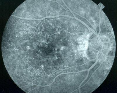
Here are two examples of what dry AMD looks like using fundus photography.
Dry macular degeneration photo

A retina with soft drusen from dry AMD.
Dry macular degeneration photo

A retina with dry AMD.
Wet AMD Fundus Photographs
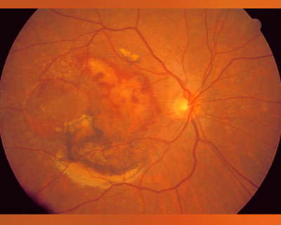
With wet AMD, abnormal blood vessels grow under the retina, leak blood or other fluids, and cause scarring. This can damage your vision quickly.
This is what wet AMD looks like using fundus photography.
Wet macular degeneration photo

A retina with abnormal, leaking blood vessels of wet AMD.
Video: Age-Related Macular Degeneration (AMD) Explainer