Blink
Cloquet’s Canal
By David L. Greenburg, MD, MPH, Walter Reed National Military Medical Center, Bethesda, Md.
Download PDF

A 52-year-old retired military officer was scheduled for removal of a moderately dense posterior subcapsular cataract. A month before, a very dense posterior subcapsular cataract had been removed uneventfully from his fellow eye; that procedure was his only ocular history.
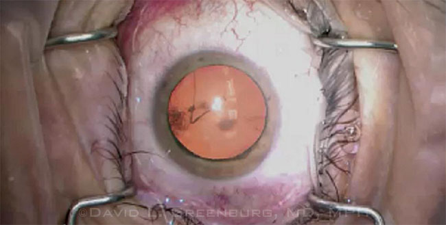
At the time of our surgery, a thin, translucent tubular reflection was noted in the vitreous just posterior to the lens capsule. Highlighted against the red reflex of the operating microscope, the structure was observed to pass posteromedially toward the optic nerve. Its anterior aspect was decentered slightly inferonasally to the posterior pole of the lens capsule. The patient’s cataract extraction progressed uneventfully. At the postoperative dilated examination, the structure was still visible posterior to the lens capsule in the vitreous. No abnormalities were noted at the optic nerve head.
During embryologic development, the hyaloidal vasculature passes through Cloquet’s canal to perfuse the embryonic lens before regressing during the second trimester. We believe the observed structure represents Cloquet’s canal.
| BLINK SUBMISSIONS: Send us your ophthalmic image and its explanation in 150-250 words. E-mail to eyenet@aao.org, fax to 415-561-8575, or mail to EyeNet Magazine, 655 Beach Street, San Francisco, CA 94109. Please note that EyeNet reserves the right to edit Blink submissions. |
Multimedia Extra
The video above takes another look at Cloquet’s canal.