By Derek Huang, MD, and Rona Z. Silkiss, MD, FACS
Edited by Sharon Fekrat, MD, and Ingrid U. Scott, MD, MPH
Download PDF
Merkel cell carcinoma of the eyelid is a rare, highly malignant neuroendocrine tumor. This type of tumor originates from sensory Merkel cells, which were discovered by Friedrich Merkel in 1875. These cells, located in the basal epidermis, are associated with perception of light touch and the discrimination of shapes and textures. When they undergo malignant transformation, Merkel cell carcinomas can arise.1 These carcinomas have a predilection for the head and neck area (representing 50 percent of cases); of greatest relevance to ophthalmologists, 5 to 10 percent of all cases occur on the eyelid. The annual incidence is 0.23 cases per 100,000 persons.2
Clinical Presentation
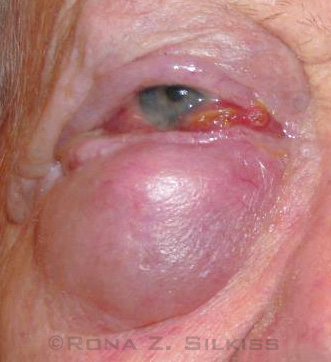
Merkel cell carcinomas can involve the upper and lower eyelid (Fig. 1), but the upper lid is the more common site at presentation. The lesion commonly presents as a painless nodule with a violaceous purple-red hue and occasional ulceration. There may be sparing or partial loss of the eyelashes. The overlying skin may develop a hard, smooth-surfaced texture that may be reddened as a result of telangiectasia. Rapid growth (usually within six months) and early lymph node metastasis generally occur.3
The mnemonic AEIOU—asymptomatic, expanding rapidly, immune suppression, older than 50 years, and ultraviolet-exposed site on a person with fair skin—has been used to describe the presentation of this type of carcinoma. In a series of 195 patients, 89 percent of patients with primary Merkel cell carcinoma had three or more of these presenting indicators.4
Possible misdiagnoses. Because of its rarity, Merkel cell carcinoma is not usually suspected clinically before biopsy, which can lead to diagnostic delay and inadequate treatment.5 It can be misdiagnosed as a cyst, chalazion, nodular angiosarcoma, keratoacanthoma, basal cell carcinoma, sebaceous carcinoma, or lymphoma.3 Clinicians should suspect Merkel cell when a lesion is rapidly growing and nodular in appearance.5 Other eyelid malignancies, such as basal cell, squamous cell, and sebaceous carcinomas, typically grow more slowly and often have signs and symptoms for more than one year before presentation.
Contributing risk factors. Merkel cell carcinoma is usually observed in individuals older than 50 years. The large majority of patients are Caucasian, with a female predominance.1 Because this carcinoma has a predilection for sun-exposed skin, a history of extensive sun exposure also increases risk. Immunocompromised patients are affected disproportionately. Recently, polyomavirus has also been implicated as a risk factor, with 80 percent of Merkel cell carcinomas testing positive for the virus.3
 |
|
AN ADVANCED CASE. This 73-year-old patient has advanced Merkel cell carcinoma involving both the upper and lower eyelids of the right eye.
|
Diagnosis and Management
When Merkel cell carcinoma is suspected, ensuring the correct diagnosis as well as delineating the extent of disease promptly is paramount in improving outcomes.
Differential diagnosis. The differential diagnosis of Merkel cell carcinoma includes chalazion, sebaceous carcinoma, squamous cell carcinoma, and basal cell carcinoma.
Biopsy and histopathologic analysis. An excisional or full-thickness incisional biopsy of the suspected lesion should be performed, followed by careful histopathologic analysis. Lesions tend to have large subepidermal nests of cells with scant cytoplasm. The cells have large nuclei that are round to oval in shape with finely dispersed chromatin, a vesicular appearance, and numerous mitotic figures.
Because Merkel cell carcinoma can be confused with sebaceous carcinoma, additional immunohistochemistry and electron microscopy can be of great value in differentiation. Merkel cell carcinoma expresses cytokeratin polypeptides 8, 18, and 19, which are characteristic of epithelia. It also exhibits a distinct marker profile with dotlike coexpression of pancytokeratin with cytokeratin 20. It stains positively for neuron-specific enolase and negatively for S-100 protein.6 Electron microscopy demonstrates dense-core cytoplasmic granules.3
Clinical examination. A complete physical examination should be performed, including a comprehensive ophthalmic exam to assess the extent of the disease. Palpation of the preauricular and cervical lymph nodes is important to assess for possible lymphatic spread. If there is any evidence of lymphadenopathy or if the eyelid appears to be diffusely involved, additional imaging is required to evaluate for systemic spread of the disease.
Management
Management consists of wide surgical excision with suggested margins of 2.5 to 3.0 cm and pathologic nodal staging.2 Mohs micrographic surgery or frozen sections are used for histopathologic confirmation of disease-free margins of 5 mm around the eyelids.3,7 Sentinel lymph node biopsy is helpful in detecting metastases that spread through the lymphatic system and aids in disease staging.
Adjuvant chemotherapy and combination treatments have been reported to be successful in limited cases for palliative purposes or inoperable disease.3 The role of adjuvant radiotherapy remains controversial, as there is inconclusive evidence for improvement in disease-specific mortality.2
Merkel cell carcinoma has a high rate of recurrence, with a reported incidence of 21 percent in one series.1
Prognosis
The estimated mortality rate for all patients with Merkel cell carcinoma is 25 to 35 percent.2 A review of 251 Merkel cell carcinoma patients found a 97 percent five-year survival rate in patients who were node negative on pathologic study; however, the five-year survival dropped to 52 percent in those with pathologically positive nodes.8
Some factors associated with a worse prognosis include male gender, which in one study portended a survival rate of 58 percent compared with 80 percent in women.3 Histologic characteristics associated with a poorer prognosis include the size of the tumor, invasion of subcutaneous fat, heavy lymphocytic infiltrate, and a high number of mitotic figures.9
Conclusion
Merkel cell carcinoma is a rare and aggressive tumor that necessitates prompt diagnosis and treatment. A high index of suspicion is important in patients who present with risk factors to avoid initial misdiagnosis and delay of therapy. Although there is no universal management protocol, a growing consensus indicates that aggressive treatment with wide surgical excision and lymph node dissection, adjuvant chemotherapy, and/or radiotherapy may help decrease the rate of recurrence and increase survival.
___________________________
1 Peters GB 3rd et al. Ophthalmology. 2001;108(9):1575-1579.
2 Herbert HM et al. JAMA Ophthalmol. 2014;132(2):197-204.
3 Barrett RV, Meyer DR. Int Ophthalmol Clin. 2009;49(4):63-75.
4 Heath M et al. J Am Acad Dermatol. 2008;58(3):375-381.
5 Colombo F et al. Ophthal Plast Reconstr Surg. 2000;16(6):453-458.
6 Furuno K et al. Jpn J Ophthalmol. 1992;36(3):348-355.
7 Pathai S et al. Orbit. 2005;24(4):273-275.
8 Allen PJ et al. J Clin Oncol. 2005;23(10):2300-2309.
9 Gess AJ, Silkiss RZ. Ophthal Plast Reconstr Surg. 2012;28(1):e11-13.
___________________________
Dr. Huang is a resident in ophthalmology, and Dr. Silkiss is chief of the Division of Ophthalmic Plastic, Reconstructive and Orbital Surgery, at California Pacific Medical Center in San Francisco. The authors report no related financial interests.