By Umesh Krishna, MD, Sumana J. Kamath, MD, and Madhurima K. Nayak, MD
Edited by Sharon Fekrat, MD, and Ingrid U. Scott, MD, MPH
Download PDF
Vitamin A deficiency (VAD) can cause a range of ocular manifestations, known collectively as xerophthalmia, including night blindness, conjunctival and corneal xerosis, and keratomalacia, and is an important cause of preventable blindness. The major cause of VAD is malnutrition, followed by malabsorption.1
The World Health Organization has classified the ocular manifestations of VAD as follows2:
XN Night blindness
X1A Conjunctival xerosis
X1B Bitot’s spots
X2 Corneal xerosis
X3A Corneal ulceration <1/3 of surface
X3B Corneal ulceration >1/3 of surface
XS Corneal scarring
XF Xerophthalmic fundus
This article will focus on X1B, Bitot’s spots.
 |
|
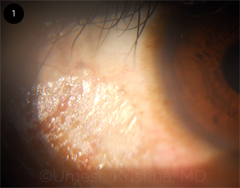
BITOT’S SPOTS. Conjunctival lesion, temporal to the cornea, shows typical dry, foamy appearance.
|
Presentation
Bitot’s spots, first described by the French physician Pierre Bitot in 1863 in debilitated children,3 are an important sign for diagnosing vitamin A deficiency (VAD). Bitot’s spots are typically dry-appearing triangular patches of xerosed conjunctiva with a layer of foam on the surface, usually located temporal to the cornea (Fig. 1). These lesions are not wetted by the tear film. Bitot’s spots are more extensive than lesions caused by focal conditions of the ocular surface such as trachoma, burns, and pemphigoid.4
Pathophysiology
Vitamin A is required for the development of normal epithelium. VAD causes Bitot’s spots through metaplasia of the conjunctival epithelium and tangles of keratin admixed with Corynebacterium xerosis, which dwell in the stratum corneum of the conjunctiva. The typical foamy appearance is due to gas produced by these bacteria.5 Histologically, Bitot’s spots show keratinization, irregular maturation, inflammatory infiltration, and accumulation of gram-positive bacilli.6
In developing countries, primary VAD is mainly caused by malnourishment,1 particularly from decreased intake of provitamin carotenoids. Pregnant and lactating women and young children are at greatest risk, owing to higher nutritional demands. Further, in children with measles, VAD increases morbidity and mortality.
In developed countries, VAD is usually secondary to gastrointestinal disorders that cause malabsorption or impaired storage or transport of vitamin A, including liver, bowel, or pancreatic disease.7 Deficiency may also occur in people following strict vegetarian or vegan diets.
Causes. The major underlying causes of VAD may be summarized as follows.8,9
Reduced intake
- Inadequate food supply
- Alcoholism
- Mental illness
- Dysphagia
Impaired absorption
- Crohn’s disease
- Celiac sprue
- Pancreatic insufficiency
- Short bowel syndrome
- Chronic diarrhea
Disordered transport
Reduced storage
- Liver disease
- Cystic fibrosis
Role of zinc. Zinc deficiency may also be involved with the pathogenesis of secondary VAD. Inadequate zinc can depress the hepatic synthesis of retinol-binding protein (RBP), which is required for mobilization of retinol from the liver. In addition, zinc may play a role in the conversion of beta-carotene to retinol via the enzyme 15-15 dioxygenase.10
Evaluation
In addition to an ocular examination, the evaluation of a patient with Bitot’s spots involves a careful history, general health exam, and laboratory investigations to determine the underlying cause (Chart 1). In patients presenting with Bitot’s spots, evaluate first for malnutrition in the younger age group and for malabsorption in the elderly.
History. The clinician should query the patient or guardian on aspects of the social or medical history potentially associated with reduced intake or impaired absorption of vitamin A. Relevant factors include age, diet, weight loss, alcohol intake, gastrointestinal disorders or surgery, and night blindness or other ocular conditions.
General exam. This should include assessment of the patient’s build and weight, signs of jaundice, and abdominal palpation to rule out hepatomegaly.
Ocular exam. The clinician should look for possible subconjunctival fibrosis and symblepharon. The status of the ocular surface may be evaluated by means of Schirmer test, rose bengal, or lissamine green staining, and conjunctival impression cytology.
Blood tests. Serum RBP measurement is relatively simple, is inexpensive, has high specificity and sensitivity, and can be done in less-advanced laboratories.11 The reference range is 30 to 75 mg/L.
Serum vitamin A/retinol. The reference range is 30 to 80 μg/dL.
Serum zinc. The reference range is 75 to 120 μg/dL.
Treatment
High-dose vitamin A is the treatment for all individuals with xerophthalmia and for infants or children with severe malnutrition or measles. See the treatment regimens in Table 1.
Improvement of Bitot’s spots is seen within 2 weeks of high-dose vitamin A therapy. However, the retinal manifestations of vitamin A deficiency are slower to respond to treatment, with night blindness and dark adaptation problems often persisting for 4 weeks.12
___________________________
1 Sharma A at al. Int J Prev Med. 2014;5(8):1058-1059.
2 Sommer A. Vitamin A Deficiency and Its Consequences. Geneva: WHO; 1995. http://apps.who.int/iris/handle/10665/40535.
3 Shukla M, Behari K. Indian J Ophthalmol. 1979;27(2):63-64.
4 Sihota R, Tandon R. Diseases of the conjunctiva. In: Parson’s Diseases of the Eye, 20th ed. New Delhi: Elsevier India; 2007:178.
5 Ferrari G et al. N Engl J Med. 2013;368(22):e29.
6 Sommer A et al. Arch Ophthalmol. 1981;99(11):2014-2027.
7 Ahad MA et al. Eye. 2003;17(5):671-673.
8 Rubino P et al. Case Rep Ophthalmol Med. 2015. doi:10.1155/2015/181267.
9 Fernando-Langit A, Ilsen PF. Clin Refract Optom. 2008;19(3):86-93.
10 Tinley CG et al. J Cyst Fibros. 2008;7(4):333-335.
11 Mahmood K et al. Saudi J Gastroenterol. 2008;14(1):7-11.
12 Ross DA. J Nutr. 2002;132902S-2906S. http://jn.nutrition.org/content/132/9/2902S.full.
___________________________
Dr. Krishna is a resident, Dr. Kamath is a professor, and Dr. Nayak is senior resident in the department of ophthalmology, Kasturba Medical College, Mangalore, Manipal University, Karnataka, India. Relevant financial disclosures: None.