Blink
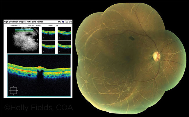
Presumed (Inactive) Ophthalmomyiasis
By Rebecca A. Manning, MD, North Carolina Retina Associates, Raleigh, N.C.
Photo by Holly Fields, COA, North Carolina Retina Associates, Raleigh, N.C.
Download PDF

A 79-year-old African-American man was referred for an incidental finding of an abnormal right fundus during cataract evaluation. In 1976, he had been told that his eye had “abnormalities,” but no specific diagnosis was made at that time. He has not had any visual complaints over the subsequent years.
He presented to us with vision of 20/25, and no active anterior or posterior inflammation. Fluorescein angiography revealed extensive areas of hyperfluorescent, linear lesions throughout the posterior pole extending into the periphery. There was a mildly elevated but inactive chorioretinal scar superior to macula. The left eye was normal. We concluded that these findings were most likely the result of ophthalmomyiasis—an infection of the eye with fly larvae—in the distant past.
No treatment is warranted, given the absence of inflammation or visual complaints.
| BLINK SUBMISSIONS: Send us your ophthalmic image and its explanation in 150-250 words. E-mail to eyenet@aao.org, fax to 415-561-8575, or mail to EyeNet Magazine, 655 Beach Street, San Francisco, CA 94109. Please note that EyeNet reserves the right to edit Blink submissions. |