Download PDF
Highlighted Paper at AAO 2015: Intraocular Inflammation, Uveitis
Sequential imaging of the choroid with swept-source optical coherence tomography (SS-OCT) can enable clinicians to monitor choroidal thickness and morphology in patients undergoing treatment for birdshot chorioretinopathy (BCR), Spanish researchers will report at AAO 2015.
During 1 year of follow-up, the group used an SSOCT system (Atlantis DRI OCT-1, Topcon) to image 24 eyes with BCR uveitis and 42 control eyes, said study coauthor Sara Jordán Cumplido, MD.
 |
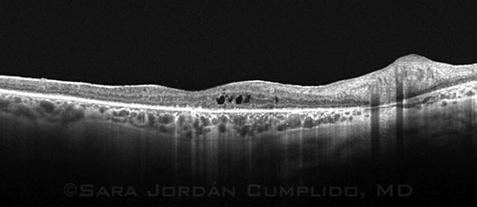
Swept-Source OCT. B-scan shows retinal and choroidal changes in a patient with BCR. Retina: intraretinal cysts, hyperreflective foci in outer layers, and generalized loss of the ellipsoid zone. Choroid: stromal fibrosis, hyperreflective foci, and suprachoroidal and hyporeflective space.
|
Capabilities of SS-OCT. The Atlantis captures up to 100,000 A-scans per second and combines the data into quantified, 3-dimensional B-scan images that can be used to examine anatomic structures at different depths inside the eye, explained Dr. Jordán Cumplido, who is an ophthalmology resident at the Hospital Universitari de Bellvitge, in Barcelona.
“The higher capabilities of SS-OCT at detecting the chorioscleral interface make it a reliable tool for measuring choroidal thickness, the mainstay of our study,” she said. “SS-OCT allows for deeper penetration into the retinochoroidal structures as well as increased resolution, while displaying a simultaneously focused image of both retina and choroid.”
| Swept-Source OCT and Active Birdshot Chorioretinopathy. When: Monday, Nov. 16, 11:45-11:52 a.m., during the intraocular inflammation, uveitis original papers session (10:45 a.m.-12:15 p.m.). Where: Bassano 2701. Access: Free. |
Imaging features of BCR. In the retina, SS-OCT showed focal loss of architecture, hyperreflective foci, and intraretinal cysts. Choroidal findings were thinning of the Sattler layer, hyperreflective foci, and increased transmission effect.
The researchers found that OCT thickness data correlated well with the results of indocyanine green angiography and visual field testing in the uveitis patients over time, Dr. Jordán Cumplido said. Mean choroidal thickness was 247.6 ± 70 μm in the BCR patients and 287.5 ± 69 μm in the controls. The choroid thinned an average of 33 μm in 4 cases with angiographic and visual field improvements; was unchanged in 7 patients with stable results; and thickened by 53 μm in 1 patient whose other test results and visual acuity worsened.
—Linda Roach
___________________________
Relevant financial disclosures—Dr. Jordán Cumplido: None.
For full disclosures and disclosure key, see below.

More from this month’s News in Review