Blink
Epithelial Ingrowth
By Gary L. Legault, MD, and Alan N. Carlson, MD, and photographed by Tiffanie Keaton, Duke Eye Center, Durham, N.C.
Download PDF

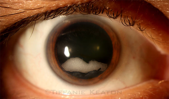
A healthy 56-year-old man who had undergone LASIK in our clinic 14 years ago returned, noting a gradual decrease in near vision over several years. The patient described a “white spot” developing on his right cornea. Vision in the right eye was 20/30 at distance and J1+ at near with a +2.00 lens. Examination revealed a white, dense, plaque-like lesion, measuring 2.0 × 6.4 mm, in the LASIK flap interface inferior to the visual axis in the right eye. The LASIK flap was clear in the left eye.
The presumed diagnosis is epithelial ingrowth. A flap lift and irrigation were performed. On postoperative day 1, the opacity was significantly clearer; however, residual scarring outlining his lesion was present.
This presentation of epithelial ingrowth is unusual because of the density and large area of ingrowth. The patient had moved away from the area shortly after his LASIK surgery without any follow-up care, and this contributed to the extent of this growth.
Epithelial ingrowth is typically seen 1 mm from the LASIK flap edge and is usually not visually significant.
| BLINK SUBMISSIONS: Send us your ophthalmic image and its explanation in 150-250 words. E-mail to eyenet@aao.org, fax to 415-561-8575, or mail to EyeNet Magazine, 655 Beach Street, San Francisco, CA 94109. Please note that EyeNet reserves the right to edit Blink submissions. |