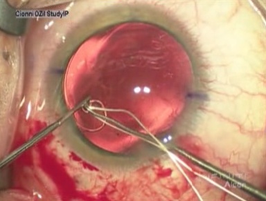
Robert J. Cionni, MD, provides an overview and demonstration of how to use the Morcher capsular tension rings in patients with severe zonular damage.
Introduction
Regardless of etiology, patients with weak or missing zonules are at higher risk of complications during and after cataract surgery. Capsular tension rings (CTRs), modified capsular tension rings (MCTRs) (see figure 1) and capsular tension segments (CTSs) do provide us a better opportunity to successfully manage these cases and allow for implantation of a PC IOL into the capsular bag.1,2,3,4,5,6,7

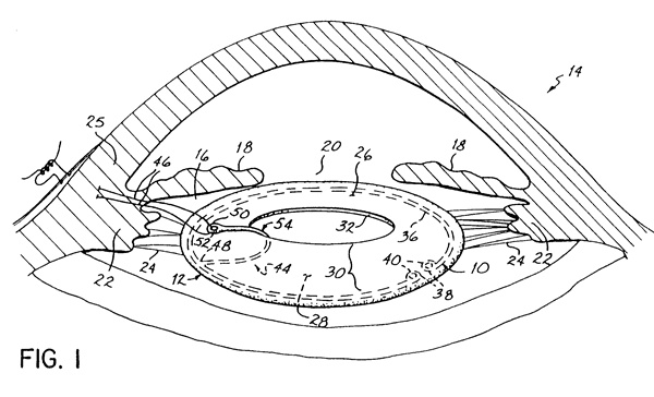
Figure 1. Modified capsular tension ring Model I-L
Eyes with profound zonular compromise (more than 180 degrees of zonular dialysis or significant lens subluxation) are unlikely to achieve adequate stabilization or centration with a standard CTR. For these patients, the MCTR provides for scleral fixation of the CTR without violating the integrity of the capsular bag; stabilizing and centering the capsular bag so that a PC IOL can be placed into the bag.
Surgical Technique
Some general concepts and a careful preop exam will help elucidate likely requirements for successful surgery. Traumatic lens subluxation typically preserves zonules that are quite strong in at least one quadrant, which allows for a single fixation hook style MCTR. But occasionally trauma can induce even more extensive zonular damage which requires a double sutured MCTR. Lens subluxation due to pseudoexfoliation syndrome can often be managed with a standard CTR but more severe cases may require a MCTR for long-term stabilization. Congenital lens subluxation behaves quite differently, characterized by either a round or flattened lens equator. A flattened or otherwise deformed lens equator indicates that the zonules attached to the flattened portion of the lens are much weaker than the zonules remote from it; therefore, a single fixation point at the site of the weakest zonules will likely be sufficient. However, when the lens is decentered and the equator perfectly round, all zonules are quite weak as they have allowed the lens capsule to contract nearly equally for 360 degrees. For these patients a double sutured MCTR is likely the best choice. Expect surgery to be significantly more challenging in these cases.

View video of Dr. Cionni using the Morcher Capsular Tension Ring
Each case begins by injecting a dispersive ophthalmic viscoelastic device (OVD) to tamponade vitreous from anterior movement. The capsulorrhexis (CCC) tear begins remote from the area of greatest weakness, using the counter traction provided by the remaining healthy zonules. The CCC is fashioned to be shaped similar to the lens since an oval lens capsule will become round once a MCTR is in place, and the CCC will conform to this shape as well. As the capsulorrhexis proceeds it may be necessary to stop and place iris hooks for counter traction. Although a MCTR can be placed immediately after CCC, I prefer to place the ring as late as possible during the procedure.8 Instead of early MCTR insertion, I expand the bag behind the lens nucleus with a dispersive OVD. Using this technique I find it rarely necessary to support the bag with any devices until the nucleus has been emulisfied and removed. Alternatively, you can stabilize the capsular bag by engaging the capsulorrhexis edge with one to three disposable nylon iris retractors placed through limbal stab incisions or placing a CTS secured it with an iris hook or scleral suture. Generous hydrodissection should be performed to maximally free the nucleus. If the epinucleus is soft enough this will cause the nucleus to prolapse into the anterior chamber.
Phacoemulsification should be performed using lower vacuum and aspiration settings to minimize chamber volatility.9 Torsional phacoemulsification is ideal for cases with zonular dehiscence as it minimizes lens material chatter, which may decrease the risk of losing nuclear material through areas of missing zonules (Cionni R, Comparison of Nuclear Material Chatter: Longitudinal Versus Torsional Phacoemulsification, Presented at: American Society of Cataract Surgery, April, 2007, San Diego, CA).
Use a dispersive OVD to "viscodissect" the nuclear halves or quadrants free from the cortex in areas of zonular weakness and lift them into a safe plane for emulsification. The OVD will expand and stablize the capsular bag while keeping the vitreous posterior to the working space.
Before inserting a CTR or MCTR, inject OVD under the surface of the anterior capsular rim to create a space for the ring and to dissect residual cortex away from the peripheral capsule, reducing the likelihood of cortical entrapment by the ring. I modified my technique for MCTR insertion following a suggestion from Michael Hater, MD, at the Cincinnati Eye Institute. A 9.0 prolene or 8.0 gortex suture is pre-placed through the eyelet of the fixation hook before inserting the ring into the capsular bag. (Note: gortex sutures for intraocular surgery is considered an "off-label" use) The MCTR, now available in a small and large size, is inserted with smooth forceps through the main incision and dialled into the capsular bag with a Sinskey hook or a Y-hook. While the ring dials into the bag, the fixation hook typically "captures" anterior to the capsulorrhexis edge. However, if it does not, the hook is easily manipulated above the capsular bag with two dull instruments. The MCTR fixation hook is then dialed to the center of the zonular dialysis. A 23-gauge MVR blade is used to fashion two scleral incisions, 2-3mm posterior to the limbus and about 2mm apart. 25-gauge retinal forceps are placed through the sclerotomy sites and the sutures are grasped and externalized. The suture is tightened to the point of bag centration and tied. Because the gortex suture is quite slippery, take care not to overtighten the suture. The knot is then rotated through the sclerotomy sites. Sclerotomies don't require sutures unless there is fluid leakage.
After suture fixation of the MCTR, any remaining cortex can be aspirated, and the capsular bag re-inflated with a cohesive OVD before PC IOL insertion. In these cases I have found that inserting a foldable-style single-piece acrylic PC IOL into the capsular bag is easiest.
Don't remove all of the OVD since doing so might encourage vitreous prolapse late in the procedure. Similarly, whenever exiting the chamber, always provide infusion through the side-port incision to prevent chamber collapse. If vitreous presents at any time during the procedure, it should be carefully and completely removed from the anterior chamber. I prefer a pars plana approach and triamcinalone staining to best assure that the anterior chamber is free of vitreous. Finally, acetylcholine is instilled at the end of the procedure to ensure that the pupil rounds which further confirms that the anterior chamber is free of vitreous.
Complications
I have seen two types of postoperative complications related to the MCTR. The first is iris chaffing due to the fixation hook being sutured too anteriorly. The chaffing is usually self limited but can lead to UGH syndrome, which would necessitate repositioning of the fixation point more posteriorly. The second complication is late suture lysis. This typically occurs when using 10.0 prolene suture but can occur with 9.0 prolene as well, years after the initial procedure. If using 8.0 gortex suture it is highly unlikely to ever occur. Management is actually quite simple. Using a double-armed gortex suture, the ring is re-sutured to the scleral wall by passing one needle through the bag, posterior to the ring and the second suture anterior to the ring and bag. The capsular bag will not "splay" open as capsular fibrosis holds it strong.
References
- Cionni RJ, Osher RH. Endocapsular ring approach to the subluxed cataractous lens. J Cataract Refract Surg. 1995;21:245-249.
- Cionni RJ, Osher RH. Management of profound zonular dialysis or weakness with a new endocapsular ring designed for scleral fixation. J Cataract Refract Surg. 1998;24:1299-1306.
- Cionni RJ, Osher RH, Marques DM, Marques FF, Snyder ME, Shapiro S. Modified capsular tension ring for patients with congenital loss of zonular support. J Cataract Refract Surg. 2003;29:1668-1673.
- Hasanee K, Butler M, Ahmed, II. Capsular tension rings and related devices: current concepts. Curr Opin Ophthalmol. 2006;17:31-41.
- Hara T, Yamada Y. "Equator ring" for maintenance of the completely circular contour of the capsular bag equator after cataract removal. Ophthalmic Surg. 1991;22:358-359.
- Nagamoto T, Bissen-Miyajima H. A ring to support the capsular bag after continuous curvilinear capsulorhexis. J Cataract Refract Surg. 1994;20:417-420.
- Legler U, Witschel B. The capsular ring: a new device for complicated cataract surgery. Film presented at: American Society of Cataract Surgery. May 1993; Seattle, Washington.
- Ahmed, II, Cionni RJ, Kranemann C, Crandall AS. Optimal timing of capsular tension ring implantation: Miyake-Apple video analysis. J Cataract Refract Surg. 2005;31:1809-1813.
- Osher RH. Slow motion phacoemulsification approach. J Cataract Refract Surg. 1993;19:667.
Author Disclosure
Dr. Cionni, as its developer, has a financial interest in the modified capsular tension ring (MCTR). He is also a consultant for Alcon Laboratories, Inc.