The links to each individual chapter of the Color Atlas of Gonioscopy are available at the Chapters link, below.
Gonioscopy is a relatively young science, having been developed entirely within the twentieth century.
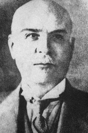
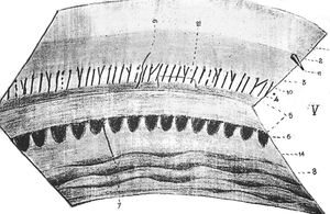
The Greek ophthalmologist Alexios Trantas (2‑1) first reported examination of the angle in 1907. He viewed the angle in a patient with keratoglobus using a direct ophthalmoscope while indenting the sclera with his finger (Trantas, 1907). Some years later he presented remarkably detailed drawings of the angle (2‑2) (Trantas, 1918). He coined the term “gonioscopy”, meaning “observation of the angle”, from the Greek (Dellaporta, 1975).
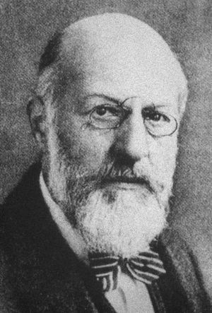
Maximilian Salzmann (2‑3) recognized that the normal angle was not visible owing to total internal reflection (Salzmann, 1914). He was the first to view the angle through a contact lens and, in 1915, presented a paper with excellent drawings of the angle obtained by means of his newly developed contact lens (Salzmann, 1915). Salzmann stressed the importance of gonioscopic examination in patients with a history of angle closure. He recognized that the development of synechiae in the angle did not always lead to elevated intraocular pressure. Salzmann was also the first to describe blood in Schlemm’s canal.

2-1 Alexios Trantas (1867–1961). (Reprinted from Survey of Ophthalmology, Vol 20, Dellaporta A, Historical notes on gonioscopy, pages 139–149, copyright 1975, with permission from Elsevier.),

2-2 This drawing of the angle, made by Trantas in 1918, demonstrates remarkable detail. The angle was viewed with an ophthalmoscope while the limbus was indented by the examiner’s finger. (Reprinted from Survey of Ophthalmology, Vol 20, Dellaporta A, Historical notes on gonioscopy, pages 139–149, copyright 1975, with permission from Elsevier.)

2-3 Maximilian Salzmann (1862–1954). (Reprinted from Survey of Ophthalmology, Vol 20, Dellaporta A, Historical notes on gonioscopy, pages 139–149, copyright 1975, with permission from Elsevier.)
Mizuo (1914) examined the inferior angle in patients by everting the lower lid and filling the cul-de-sac with saline. The technique was difficult to perform because the saline lens was lost when the patient blinked.
The introduction of Zeiss’ slit lamp permitted significant advances in gonioscopy. Koeppe (1919) used the Zeiss slit lamp to examine the angle with his newly developed lens, which was thicker and more convex than Salzmann’s lens. Gonioscopy was performed with the patient seated at the slit lamp. A knotted bandage rested on a central depression in the lens to secure it to the patient. This technique was effective only for evaluating the nasal and temporal sectors of the angle.
In 1925 Manuel Uribe Troncoso developed a self-illuminating monocular gonioscope that permitted examination of all parts of the angle (Troncoso, 1925).
Thorburn was the first to photograph the angle. In 1927 he photographed an instance of angle closure brought on by mydriatics and subsequently reversed by eserine. He also observed that the majority of his patients with glaucoma had open angles (Thorburn, 1927).
Otto Barkan used a slit lamp suspended from the ceiling and a hand-held illuminator to view the angle through a Koeppe lens (Barkan et al, 1936). His technique had the advantage of bright illumination and sufficient magnification, and his apparatus brought gonioscopy into practical clinical application. He subsequently made the distinction between “deep- chamber” and “ shallow-chamber” glaucoma, and suggested that iridectomy be used for shallow-chamber glaucoma only (Barkan, 1938).
Barkan was also the first to describe goniotomy under direct visualization (Barkan, 1937).
Indirect gonioscopy was introduced with the Goldmann mirrored contact lens (Goldmann, 1938). The Allen lens, developed a few years later, used a totally refractive prism rather than a mirror (Allen and O’Brien, 1945). This was later modified into the Allen-Thorpe gonioprism, which had four prisms and permitted most of the angle to be viewed without rotation of the lens (Allen et al, 1954).
The first attempt to grade the angle was that of Gradle and Sugar (1940). Scheie (1957) developed a grading system based on visible structures. The widely used Shaffer grading technique was developed 3 years later (Shaffer, 1960). Spaeth modified this system to provide information regarding the angle of iris approach, the level of iris insertion, and the configuration of the iris (Spaeth, 1971). The techniques of angle grading are described more completely in Chapter 6.
An excellent review of the history of gonioscopy has been provided by Dellaporta (1975).