This case study examines the biological cause of anterior lens iridescence present in individuals with a rare hereditary condition. The 59-year-old patient with polychromasia capsulare belongs to one of the 3 families in which the autosomal dominant trait is known to run.
The patient presented with bilateral cataract, long-standing alternating exotropia, gradual reduction in vision and diabetes mellitus. Upon preoperative biomicroscope inspection, the patient’s anterior lens capsules showed a remarkable, multi-colored array that changed with the angle of direct illumination. This display of iridescence is consistent with structural color, a mechanism found throughout the animal kingdom responsible for the vibrancy of butterfly wings, bird feathers, and the sheen of metallic beetles, among many more phenomena. Dr. Robert Osher further explains the condition in this video.
Cataract surgery was performed, and the patient consented to histopathologic examination of the anterior lens portion that was to be excised. A continuous capsulorhexis (removed by bent cystotome) measuring just under 6.0 mm was removed, divided into fragments, and sent to multiple laboratories for investigation. The cataract surgery itself was uneventful, and the patient experienced good correction in vision with a +18.5 acrylic IOL. Three months later, the patient underwent successful cataract surgery in the fellow eye, this time utilizing a femtosecond laser for capsulorhexis. This sample was also sent to the same laboratories for analysis.
After surgery, iridescent coloration was still noted on slit-beam exam on the remaining peripheral anterior capsule. Residual epithelial cells were attached to the capsule, but these were not to be responsible for the effect as it was observed across the sample in areas with and without cells.
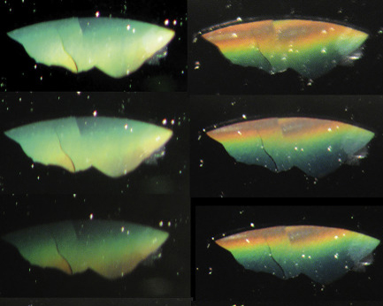
Digital macrophotography of a flat lens fragment documents the rainbow-like range of color seen as the angle of the light source is moved. When mounted flat on a standard light microscope slide and observed with direct light, the lens appeared transparent, with no iridescence.
Transmission electron microscopy (TEM) revealed an irregular polygonal structure with periodicity of 400-500 nm, compared to the uniformly amorphous makeup of a control sample. The authors sent the TEM photographs to a structural coloration expert, who concurs that this novel nanostructure is the cause of the iridescence.
Human tissue normally utilizes only primary coloration, wherein color is created by the absorption of light by pigment molecules. Individuals with polychromasia capsulare are unique in their display of structural color in the lens, and seem to present no other complications or systemic disease as a result of the condition.
