The links to each individual chapter of the Color Atlas of Gonioscopy are available at the Chapters link, below.
Grading systems permit the recording of gonioscopic findings for communication and for future reference. Several systems for grading the angle have been proposed. Gradle and Sugar (1940) quantified the depth of the angle through a Koeppe lens. They measured the distance from the plane of the iris to Schwalbe’s line with a graticule that was etched in the oculars of the microscope. Some grading systems are quite simple—for example the “wide, intermediate, and narrow” classification of Gorin and Posner (1967). The three primary alphanumeric systems that are currently used for grading the angle are those of Scheie, Shaffer, and Spaeth
Scheie System
Scheie (1957) developed a grading system in which Roman numerals were used to describe the degree of angle closure. In his system one determines the angle structures that are visible on gonioscopy (6‑1). Larger numbers signify a narrower angle. The term “wide” was used to describe an angle in which all structures were visible. Scheie also described angle pigmentation on a scale from 0 (no pigmentation) to IV (heavy pigmentation) (6‑2).
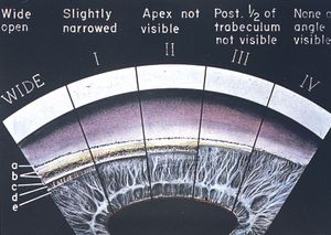
6-1 The Scheie angle depth system based on visible structures. (a, Schwalbe’s line; b, anterior and posterior trabecular meshwork; c, scleral spur; d, ciliary body face; e, iris root). (Reprinted with permission. Arch Ophthalmol. 1957;58:510– 512. Copyright © 1957, American Medical Association. All Rights Reserved.)
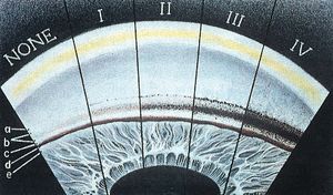
6-2 Scheie’s system of grading angle pigmentation. Larger numbers represent increasing amounts of pigmentation. Letters a through e are defined in the legend for 6‑1. (Reprinted with permission. Arch Ophthalmol. 1957;58:510–512. Copyright © 1957, American Medical Association. All Rights Reserved.)
Shaffer System
A more commonly used grading system is that of Shaffer (1960, 1962). This system describes the degree to which the angle is open rather than the degree to which it is closed. Whereas Scheie’s grade IV denotes a closed angle, on the Shaffer scale grade 4 refers to a wide-open angle. The Shaffer system approximates the angle at which the iris inserts relative to the trabecular meshwork (6‑3). If the angle between the iris and the meshwork is 20° to 45°, the angle is felt to be at no risk of closure. Angles from 0° to 20° are considered capable of closure (Table 1).
Many ophthalmologists use a variation of Shaffer’s system in which 4 is wide open and 0 is closed, but the determination is usually a rough estimate of angle width that is not strictly based on degrees.
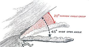
6-3 Shaffer’s grading system is based on the angle between the iris and the trabecular meshwork. For angles of 20º or less angle closure is possible. (Reprinted with permission from Trans Am Acad Ophthalmol Otolaryngol [1960;64:112– 127]. Shaffer RN. Primary glaucomas. Gonioscopy, ophthalmoscopy and perimetry.)
Spaeth System
Spaeth considered that available grading systems provided limited information and proposed a system that grades the three major features of the angle’s anatomy: the level of iris insertion, the width of the angle, and the configuration of the iris (Spaeth, 1971).
The level of iris insertion is represented by letters “A” through “E.” If the iris inserts anterior to Schwalbe’s line, it is described as grade “A” (for “anterior”). If it inserts anterior to the posterior limit of the trabecular meshwork, it is grade “B” (for “behind” Schwalbe’s line). If insertion is posterior to the scleral spur, the iris is grade “C” (for the “c” in sclera). Insertion into the ciliary body face is recorded as grade “D” (for “deep”) or grade “E” (for “extremely” deep) (6‑4).
Angular width is the estimated angle between a line tangential to the trabecular meshwork and a line tangential to the surface of the iris about one-third of the way from the periphery. The angle is expressed in degrees (6‑5).
The third characteristic that is described is the curvature of the peripheral iris: “r” for a regular or flat configuration, “s” for a steep curvature or iris bombé, and “q” for a “queer” or concave curvature (6‑6). This system was subsequently modified in order to be more descriptive, using “f ” to denote a flat configuration, “c” to describe the concave or back-bowed iris, “b” to describe the forwardly bowed iris, and “p” for a plateau iris configuration.
Spaeth graded posterior pigmented meshwork in the 12 o’clock angle on a scale of 0 to 4+. He also graded the type and number of iris processes.
The Spaeth system permits the inclusion of information obtained by indentation gonioscopy. If indentation demonstrates that the insertion is a “D” when it originally appeared to be a “C”, this would be indicated as “(C)D”. Therefore an angle is carefully defined by an alphanumeric description—such as (C)D30S—as is illustrated in 6‑7 (Spaeth, 1977).
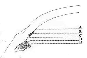
6-4 Spaeth’s classification system includes the level of iris insertion. A, anterior to trabecular meshwork; B, behind Schwalbe’s line; C, posterior to scleral spur; D, deep, into ciliary body face; E, extremely deep. (Reprinted with permission from Macmillan Publishers Ltd. Spaeth GL. The normal development of the human anterior chamber angle: a new system of descriptive grading. Eye. 1971;91:709–739.)
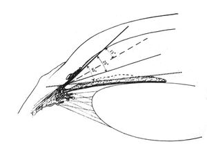
6-5 In Spaeth’s classification the width of the angle is approximated by a line tangential to the iris about one-third of the way from the iris root to the pupil and a line tangential to the face of the trabecular meshwork. (Reprinted with permission from Macmillan Publishers Ltd. Spaeth GL. The normal development of the human anterior chamber angle: a new system of descriptive grading. Eye. 1971;91:709–739.)
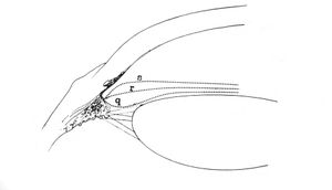
6-6 Iris configuration in the Spaeth classification. s, steep or convex; r, regular or flat; q, queer or concave. (Reprinted with permission from Macmillan Publishers Ltd. Spaeth GL. The normal development of the human anterior chamber angle: a new system of descriptive grading. Eye. 1971;91:709–739.)
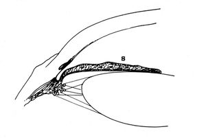
6-7 A (C)D30S angle in the Spaeth classification. The D means that iris insertion is into the ciliary body face, but the C means that the steep approach made the insertion appear to be just posterior to the scleral spur until indentation gonioscopy was performed. The number 30 is the angular width of the angle. The letter S means that the iris has a steep or convex configuration. This is an angle capable of closure. (Reprinted with permission from Macmillan Publishers Ltd. Spaeth GL. The normal development of the human anterior chamber angle: a new system of descriptive grading. Eye. 1971;91:709–739.)
Becker Goniogram
Becker (1972) described the goniogram (6‑8) as a means of drawing gonioscopic findings. He felt that this would allow description of the variable anatomy of an angle within a quadrant. It also provides a convenient way to record synechiae, tumors, foreign bodies, and so on.
Van Herick System
Mention should be made of the van Herick method of using the slit lamp to estimate the width of the angle (Table 2) (van Herick et al, 1969). A narrow slit beam is placed perpendicular to the most peripheral part of the cornea. The oculars are adjusted to give a view at an angle of about 60° from the light beam. The depth of the anterior chamber is graded by comparison to the thickness of the cornea. If the anterior chamber is thicker than the cornea, the angle is a wide-open grade 4 (6‑9). If the thickness of visible aqueous is one-quarter of the corneal thickness or less, the angle is dangerously narrow, or “slit” (6‑10).
Van Herick devised the system because routine Koeppe gonioscopy was felt to be impractical; gonioscopy was undertaken only if the angles were thought to be narrowed. Because slit-lamp gonioscopy can be rapidly performed on almost any patient, the van Herick estimation should not be relied upon as a replacement for gonioscopy. The van Herick test can be helpful in the evaluation of confusing angles because it augments gonioscopic findings by giving a separate indication of the depth of an angle. However, the test does not provide any information about the angle except depth. One could miss tumors, foreign bodies, synechiae, neovascularization, and a host of other pathologies by relying on the van Herick test alone.
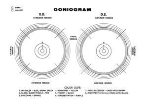
6-8 Becker’s goniogram, on which gonioscopic findings are drawn. The central dark line represents the scleral spur. The three lines outside the dark line represent the trabecular meshwork. The three lines inside the dark line represent the various levels of iris insertion into the ciliary body. A color code is provided for recording findings. (Courtesy of Stanley C. Becker, MD.)

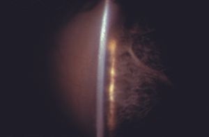
6-9 Angle estimation with the slit lamp by the van Herick method. The beam of the slit lamp is inclined at 60º from the oculars and is placed on the most peripheral cornea. The depth of the anterior chamber is compared to the thickness of the cornea. The depth of this chamber (arrow) is equal to or greater than the corneal thickness and is classified as grade 4.
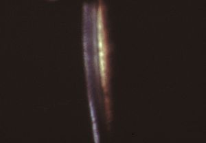
6-10 By van Herick testing this angle is narrowed to a slit and the anterior chamber is barely visible between cornea and iris. This is an angle capable of closure.
Video Clip: Gonioscopy: Van Herick Technique