Age-related macular degeneration (AMD) is a problem with your retina. It happens when a part of the retina called the macula is damaged.
In this article:
With AMD you lose your central vision. You cannot see fine details, whether you are looking at something close or far. But your peripheral (side) vision will still be normal. For instance, imagine you are looking at a clock with hands. With AMD, you might see the clock’s numbers but not the hands.
AMD is very common. It is a leading cause of vision loss in people 50 years or older.
Video: How the Eye Works and AMD
Two Types of AMD
Dry AMD
This form is quite common. About 80% (8 out of 10) of people who have AMD have the dry form. Dry AMD is when parts of the macula get thinner with age and tiny clumps of protein called drusen grow. People with dry AMD may have drusen, pigment abnormalities, or geographic atrophy (an area of cell loss in the retina). You slowly lose central vision.
Video: What Is Dry Age-Related Macular Degeneration?
Wet AMD
This form is less common but much more serious. Wet AMD is when new, abnormal blood vessels grow under the retina. These vessels may leak blood or other fluids, causing scarring of the macula. You lose vision faster with wet AMD than with dry AMD.
Many people don’t realize they have AMD until their vision is very blurry. This is why it is important to have regular visits to an ophthalmologist. They can look for early signs of AMD before you have any vision problems.
Who Is at Risk for AMD?
You are more likely to develop AMD if you:
Having heart disease is another risk factor for AMD, as is having high cholesterol levels. Caucasians (white people) also have an elevated risk of getting AMD.
Age-Related Macular Degeneration Diagnosis
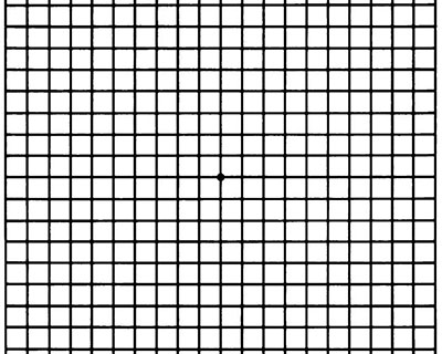
During an eye exam, your ophthalmologist may ask you to look at an Amsler grid. This grid helps you notice any blurry, distorted, or blank spots in your field of vision. Your ophthalmologist will also look inside your eye through a special lens. He or she can see if there are changes in the retina and macula.
Your ophthalmologist will put dilating eye drops in your eye to widen your pupil. This allows him or her to look through a special lens at the inside of your eye.
Optical coherence tomography (OCT) is another way to look closely at the retina. A machine scans the retina and provides very detailed images of the retina and macula.
Your doctor may do fluorescein angiography to see what is happening with your retina. Yellow dye (called fluorescein) is injected into a vein, usually in your arm. The dye travels through your blood vessels. A special camera takes photos of the retina as the dye travels throughout its blood vessels. This shows if abnormal new blood vessels are growing under the retina.
Video: Fluorescein Angiography for AMD
Optical coherence tomography angiography (OCTA) is another way to look closely at the blood vessels in and under the retina. This is like fluorescein angiography but does not use a dye.
Age-Related Macular Degeneration Treatment
Dry AMD treatment
Right now, only dry AMD with geographic atrophy can be treated. Two new medications are available: pegcetacoplan and avacincaptad pegol. One of these will be injected into your eye to slow the atrophy. Hopefully, this will delay vision loss.
There is no treatment for drusen. However people with lots of drusen or serious vision loss might benefit from taking a certain combination of nutritional supplements. A large study (AREDS and the later AREDS 2 study) found people with certain drusen may slow their dry AMD by taking these vitamins and minerals daily:
- Vitamin C (500 mg)
- Vitamin E (400 IU)
- Lutein (10 mg)
- Zeaxanthin (2 mg)
- Zinc (80 mg)
- Copper (2 mg)
Your ophthalmologist can tell you if vitamins and minerals are recommended for your dry AMD, as not all forms will benefit from the AREDS supplements. Beta carotene should not be used by smokers as it raised the risk of lung cancer.
Eye-healthy foods
Dark leafy greens, yellow fruits and vegetables, fish, and a balanced, nutrient-rich diet have been shown beneficial for people with AMD.
Wet AMD treatment
To help treat wet AMD, there are medications called anti-VEGF drugs. Anti-VEGF treatment helps reduce the number of abnormal blood vessels in your retina. It also slows any leaking from blood vessels. This medicine is delivered to your eye through a very slender needle.
Laser surgery may also be used to treat some types of wet AMD. Your eye surgeon shines a laser light beam on the abnormal blood vessels. This reduces the number of vessels and slows their leaking.
Video: Laser Therapy for Wet AMD
Talk with your ophthalmologist about ways to treat your AMD.
Making the Most of the Vision You Have
If you have AMD, you can learn how to make the most of your vision. Often you can still do many of your favorite things with special low vision tools. These can include different kinds of magnifying tools, handheld computers, electronic items and more.
Also, you can learn how to use your side vision to help you do things. A vision rehabilitation specialist can teach you how this works. They also can help you find many low vision support services and tools.
Ask your ophthalmologist to help you find a vision rehabilitation specialist in your area. The goal is to learn new ways to be as independent as possible.
Test Your Vision with the Amsler Grid
AMD causes your vision to change over time. You may not notice these changes when they happen. But you need to catch vision changes as soon as possible. Treating them early can help slow or stop further loss of sight.
You should use an Amsler grid every day to monitor your vision. One is below for you to use.

Here is how to use the Amsler grid:
- Keep the Amsler grid in a place where you see it every day. Many people keep an Amsler grid on their refrigerator door or on their bathroom mirror.
- In good light, look at the grid from about 12 to 15 inches away. Be sure to wear your reading glasses if you normally use them.
- Cover one eye. Look directly at the dot in the center of the grid with your uncovered eye. Notice if any of the lines look bent or wavy. See if any part of the grid looks blurry, dim, or out of shape.
- Now cover your other eye and test your vision this same way again.
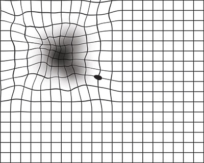
Call your ophthalmologist right away if you notice that any lines or parts of the grid look wavy, blurry, or dim.

This is what an Amsler grid might look like with blurry, wavy lines, or dim areas.