What is visual field testing?
As you focus on the words in this article, how much can you see out of the corners of your eyes? Can you tell what's happening in your surroundings?
Your visual field is how wide of an area your eye can see when you focus on a central point. Visual field testing is one way your ophthalmologist measures how much vision you have in either eye, and how much vision loss may have occurred over time.
Visual field testing can detect blind spots
A visual field test can determine if you have blind spots (called scotoma) in your vision and where they are. A scotoma’s size and shape can show how eye disease or a brain disorder is affecting your vision. For example, if you have glaucoma, this test helps to show any possible side (peripheral) vision loss from this disease.
Ophthalmologists also use visual field tests to assess how vision may be limited by eyelid problems such as ptosis and droopy eyelids.
Six types of visual field tests
1. Confrontation visual field test
A common way for your doctor to screen for any problems in your visual field is with a confrontation visual field test. You will be asked to look directly at an object in front of you, (such as the doctor’s nose) while one of your eyes is covered. Your doctor may hold up different numbers of fingers in areas of your peripheral (side) vision field and ask how many you see as you look at the target in front of you.
2. Automated static perimetry test
To check for a suspected eye problem or monitor the progress of an eye disease, your ophthalmologist will rely on more specific tests to measure how you see objects in your field of vision. The automated static perimetry test is used for this purpose. It helps create a more detailed map of where you can and can’t see.
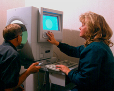
To do this test, you will look into the center of a bowl-shaped instrument called a perimeter. The eye not being tested will be covered with a patch. The testing eye will have your lens prescription placed in front of it to make sure you are seeing as well as possible.

You will be asked to keep looking at a center target throughout the test. Small, dim lights will begin to appear in different places throughout the bowl, and you will press a button whenever you see a light. The machine tracks which lights you did not see.
You may blink normally during the test. You may also pause the test if you feel you need to take a break for a moment.
Because you are looking straight ahead during the test, your doctor can tell which lights you see outside of your central area of vision. Since glaucoma affects peripheral vision, this test helps show if there is vision loss outside of your central visual field.
This is called a "static" test because the lights do not move across the screen, but blink at each location with differing amounts of brightness. This allows the machine to find the dimmest light you can see at each location in your peripheral vision.
The machine will show some lights that are too dim for you to see. This is done deliberately to find what is called the “visual threshold” of each location, meaning the brightness that you have trouble seeing half the time. You may be concerned because you can’t see every light. Rest assured that this is how the test is supposed to work.

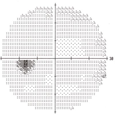
A printout of normal visual field results.
3. Kinetic visual field test
In some cases, you may have a test called kinetic visual field testing. While it is similar to the perimetry testing process described above, the kinetic test uses moving light targets instead of blinking lights.
4. Frequency doubling perimetry
Another way your ophthalmologist can assess loss of vision is using something called frequency doubling perimetry. It uses an optical illusion to check for damage to vision. Vertical bars (usually black and white) appear on the perimeter screen. These bars will flicker at varying rates. If you are not able to see the vertical bars at certain times during the test, it could show vision loss in certain parts of your visual field.
5. Electroretinography
To check for visual field loss from certain retina conditions, your ophthalmologist may also use electroretinography (ERG). This test measures the electrical signals of light-sensitive cells in the retina called photoreceptors as well as other cells. To do this test, your eyes are dilated and you will also be given numbing eye drops. Your eyes are held open with instrument called a speculum. A tiny device called an electrode is placed on your cornea. You will look into a bowl-shaped machine at flashing or varying patterns of light. The electrode measures your eye’s electrical activity in response to the light.
6. Amsler grid: A basic visual field test for central vision
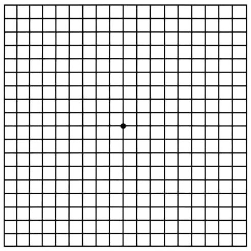
People who have age-related macular degeneration (AMD) are familiar with one very basic type of visual field test: the Amsler grid. It is a pattern of straight lines that makes a grid of many equal squares. You look at a dot in the middle of the grid and describe any areas that may appear wavy, blurry or blank.

Amsler Grid
The Amsler grid is commonly used at home by people with AMD. This test only measures the middle of the visual field, but is a simple yet helpful tool for monitoring vision changes.
How do you know if you need visual field testing?
Visual field testing is an important part of regular eye care for people who are at risk for vision loss from disease and other problems.
People with the following conditions should be monitored regularly by their ophthalmologist, who will determine how often visual field testing is needed:
- Glaucoma
- Multiple sclerosis
- Thyroid eye disease (Graves' disease)
- Pituitary gland disorders
- Central nervous system problems (such as a tumor that may be pressing on visual parts of the brain)
- Stroke
- Long-term use of certain medications (such as Plaquenil, or hydroxychloroquine, which requires yearly visual field checkups)
People with diabetes and high blood pressure have a greater risk of developing blocked blood vessels in the optic nerve and retina. They may need visual field testing to monitor any effects of these conditions on their vision.
If your visual field is limited, your ability to drive may be in jeopardy. If you are concerned about vision loss or your ability to continue driving, talk with your ophthalmologist.