Matthew Dinsdale “Dinny” Davis, MD, is best known for his key role in developing and conducting the landmark Diabetic Retinopathy Study (DRS); however, he believes that his role in recruiting outstanding people for the ophthalmology department at the University of Wisconsin Medical School in Madison may be equally, or perhaps even more, important. His greatest motivating force? His desire for the department to become the very best it could be.
“Over the years, I’ve learned to do the best that I can in every endeavor and to accept that success is often partial,” he said. “It’s been sort of a life lesson.” Dr. Davis attributes this lesson to his firstborn son, Matthew. “He had Down syndrome, and he was greatly loved by our family. One of the most important things I learned in life was from him: It was to do the best you can within your limits, to accept what ability you have, and to be forgiving of yourself.”
Dr. Davis receives the Laureate Recognition Award during the Opening Session, which takes place Sunday, 8:30-10:00 a.m., in North, Hall B.
Early Years
Dr. Davis was immersed in ophthalmology from the outset. His father, Frederick Allison Davis, MD, practiced in Madison and was the inaugural chair of the ophthalmology division in the surgery department of the University of Wisconsin Medical School in 1925. “My father was eager to have my older brother, Frederick Jefferson Davis, and me follow in his footsteps. He loved his profession, particularly the small part of his time that he devoted to ophthalmic pathology, but he refrained from being too directive,” Dr. Davis said. “By nature, I was interested in many things and reluctant to choose one too soon. So, why not choose ophthalmology, a specialty that kept many options open, included research and/or clinical practice, and allowed for the use of both medical and surgical approaches, while pleasing my father as well?”
Dr. Davis proceeded to earn his medical degree at the University of Pennsylvania. He then completed an internship and residency at the University of Wisconsin Hospital in Madison, although his residency was interrupted by 2 years of active duty in the U.S. Naval Reserve during the Korean War. When he returned, he went to Boston to finish coursework for his residency and had an extra 6 months to fill. “One of my father’s friends, Trygve Gunderson, was practicing ophthalmology in Boston, and he told my father that the latest, most fascinating thing in the field was Charles Schepens’ retina program. I spoke to Dr. Schepens, and he was kind enough to accept me for a 6-month fellowship at the Massachusetts Eye and Ear Infirmary, even though most of his fellows then came for a year. That’s how I got into retina.”
Fellowship With Dr. Schepens
Dr. Schepens, known as the founder of modern retinal surgery, was a pivotal force in Dr. Davis’ professional development. “Dr. Schepens was very kind and gentle, and I learned perseverance in spades from him,” he said. “Putting on his binocular indirect ophthalmoscope and getting to see the far peripheral retina with stereopsis and with scleral depression—which just meant pushing a little bit on the eye through the eyelid and bringing the peripheral retina into view and elevating it a little bit—was like being able to palpate an abdomen and, at the same time, seeing into the abdomen. You were seeing into the eye, and you could also palpate it. It was, and still is, absolutely amazing. It was clearly a huge breakthrough.”
During the fellowship, Dr. Schepens taught him the scleral buckling technique, which, although considered radical at the time, soon became the standard treatment for retinal detachment. Dr. Davis completed the fellowship in 1956. He then returned to Madison and joined his father, brother, and their colleagues in ophthalmic practice and started as a part-time faculty member in ophthalmology at the University of Wisconsin Medical School. “Initially, many of my retinal detachment patients were referred for reoperation after failure of an initial operation elsewhere,” he said. “Some were very challenging, and I was very thankful for all that Dr. Schepens had taught me.”
Ophthalmology at the University of Wisconsin Medical School
Peter A. Duehr, MD, who trained and practiced with Dr. Davis’ father, was chairman of the ophthalmology division at the time that Dr. Davis returned to Wisconsin. “Dr. Duehr and I both wanted ophthalmology to develop into an outstanding department, but funds and space were very limited and medical school politics were complicated. We were able to persuade several graduates from our residency program to obtain subspecialty training elsewhere and return to our division as part- or full-time faculty members. Some of them have become national leaders in their subspecialties.”
In 1970, the division achieved the status of an independent department, and Dr. Davis served as its first full-time chairman from 1970 to 1986. “Now we were able to recruit nationally. My goal was to find people who looked promising and to give them broad latitude in dividing their time between clinical practice and research. We attracted some very good people during my term as chair, and my successors have continued to do so,” he said. “Perhaps the challenge that has been my principal focus throughout my career has been to try to help make ophthalmology at the University of Wisconsin the best it can be.”
The DRS: A Landmark Clinical Trial
Dr. Davis is best known for his seminal work in diabetic retinopathy clinical trials, beginning with his position as the national chair for the DRS. At that time, approximately half of patients diagnosed with proliferative diabetic retinopathy would be legally blind within 5 years. Over the next few decades, that percentage dropped to 5%, and the DRS was a critical component in that success.
When the DRS began in 1971, it was the first major clinical trial funded by the National Eye Institute (NEI). In 1976, Dr. Davis and his collaborators published a pivotal paper based on the DRS findings, showing the substantial benefit of scatter laser photocoagulation in treating diabetic retinopathy. This finding was revolutionary because it ran counter to prevailing clinical opinion. Dr. Davis also chaired the follow-up trial, the Diabetic Retinopathy Vitrectomy Study, which demonstrated that vision was significantly better for some patients with very severe diabetic retinopathy if they had early vitrectomy surgery, as opposed to deferred surgery. These trials created standard of care treatments that are still used, and they stand as models of clinical research.
“Four decades and many clinical trials later, I am impressed with how fortunate we were to have Dinny as the chairman of the first major multicenter clinical trial in ophthalmology,” said Frederick L. Ferris III, MD, director of the Division of Epidemiology and Clinical Applications and the clinical director at the NEI. Dr. Ferris noted that “there was much skepticism about whether a clinical trial could be done in a disease with such variable outcomes and about the idea of doing multicenter randomized clinical trials at all. Many thought that careful individual physician follow-up of treated patients with reports on cohort outcomes was the better approach. There was general skepticism about whether it was possible to use standard photography and a reading center.”
But, as Dr. Ferris explained, “Dinny had the perfect collaborative personality to make things work—he is a great listener and leader. His insistence on focusing on the results of the clinical trial and letting the community develop standard practices based largely on those results, but also on clinical necessities, was brilliant. He is my model for the perfect study chairman.”
Dr. Davis said, “I had all kinds of people to help, such as Fred Ederer and Rick Ferris at the NEI, Genell Knatterud and Chris Klimt at the coordinating center, and all of the ophthalmologists and other personnel at the clinical centers. I got in on the ground floor because during the late 1960s, my colleagues and I at the University of Wisconsin had documented the natural course of diabetic retinopathy, first in serial color-coded retinal diagrams drawn by Yvonne Magli, our medical illustrator, using binocular ophthalmoscopy and later, when the Zeiss fundus camera became available to us, in stereo photographs. Our findings, particularly when presented as scientific exhibits, were so impressive that I was asked to help with this trial.”
Dr. Davis emphasized that the DRS was a joint effort. “You may notice that almost all of the DRS papers are authored by the DRS Research Group. We didn’t want any one name on them because we wanted the whole group to get equal credit. It was mainly a group effort, and the other members of the group have not gotten enough credit.”

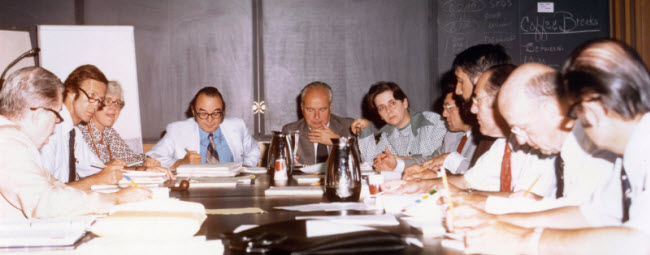
DRIVING THE DIABETIC RETINOPATHY STUDY (DRS). According to Dr. Ferris, Dr. Davis (second from the left, at a Data Monitoring Committee meeting) “was able to develop consensus among the large and diverse DRS Research Group. This was particularly important because we were creating logistics and methods for clinical trials, most of which are routine today but were new then. This involved developing the multiple committee infrastructure, including the Data Monitoring Committee and Policy Advisor Groups, where reaching consensus was critical prior to publishing results.”
The Fundus Photograph Reading Center
To grade retinal photographs collected in the DRS trial, Dr. Davis established the University of Wisconsin Fundus Photograph Reading Center (FPRC) in 1970—the first centralized, independent reading center for randomized clinical trials of retinal diseases. At the center, Dr. Davis and his collaborators developed photographic standards and systems for analyzing the characteristics of the lens and retina, and they designed quality-control systems to increase accuracy and assess reproducibility. To this day, FPRC works with researchers from around the world to analyze photographs of the retina and assess changes over time.
Classification of Diabetic Retinopathy and AMD
Finally, Dr. Davis and his collaborators developed the modified Airlie House classification of diabetic retinopathy and, later, the Early Treatment Diabetic Retinopathy Study severity scale, as well as the Age-related Eye Disease Study scales for age-related macular degeneration. “Dr. Davis is well known for his careful and complete data analysis,” said George B. Bartley, MD, editor-in-chief of Ophthalmology and soon-to-be CEO of the American Board of Ophthalmology. “These classifications remain the gold standards not only for studying these disorders but also for providing direct patient care around the world.”
Despite the extraordinary amount of hard work and dedication that Dr. Davis put into his career and the profession, he believes that he owes much to being in the right place at the right time. “A lot of luck was involved, from being in Boston in 1955 to the fact that my father knew Trygve Gunderson and asked for his advice—that was the biggest stroke of luck I ever had,” he said. “More luck arrived when Ms. Magli, a graduate in fine arts who applied for a secretarial job, rapidly mastered binocular indirect ophthalmoscopy and stereo fundus photography and joined me in following patients with diabetic retinopathy early in my career when I had time to do it.” He also considers himself extremely fortunate to have met the collaborators he worked with. “My mentors were my father and his partner, Peter Duehr, as well as Charles Schepens. From them I learned many lessons. Perhaps the most important were gentleness and perseverance.”