Blink
Can You Guess August's Mystery Condition?
Download PDF
Make your diagnosis in the comments, and look for the answer in next month’s Blink.

Last Month’s Blink
Central Cyst of the Iris Pigment Epithelium
Written by Paul M. Bell, MD, LT, MC, USN, Naval Medical Center, Portsmouth, Va., and Joseph W. Schmitz, MD, LCDR, MC, USN, Naval Medical Center, San Diego. Photo by Dr. Schmitz.

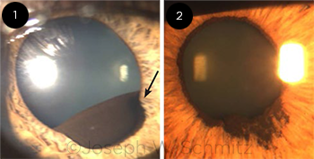
A 35-year-old man was referred by an optometrist because of a suspected iris neoplasm (Fig 1, arrow) visible in his left eye. The patient had noted an enlarging mass during the previous 2 months and decreased inferior vision in his left eye, especially when he was outside in the sunlight. He had a history of HLA (human leukocyte antigen)-B27–associated iritis in his right eye only. Dilated exam of his left eye revealed a 6.5 mm × 4.0 mm smooth, dark brown cyst on the inferior temporal margin of his iris with trace pigment cells in his anterior chamber. Of note, a small cyst was also identified under the margin of his right iris when it was fully dilated. His intraocular pressures were within the normal range and symmetrical. Gonioscopy showed an irregular contour consistent with multiple posterior masses, and the ciliary body band was visible between the elevations for ≥ 180 degrees in each eye. His visual field (VF) revealed an inferior arcuate defect in the left eye.
This is a case of a central iris pigment epithelial (IPE) cyst with a resulting VF defect. IPE cysts are uncommon, usually bilateral, and benign, and they are often identified on routine exams. Treatment is only needed in cases that are symptomatic or that cause angle-closure glaucoma or pigment dispersion syndrome. Due to this patient’s visual defect, we offered argon laser therapy, which was completed without complications. The cyst partially collapsed immediately, and the patient’s visual symptoms resolved after 2 days. One month later, (Fig. 2) the cyst was completely gone with only residual pigmentation on the iris as evidence of its prior existence. There has been no recurrence in 6 years of follow-up.
Read your colleagues’ discussion.
| BLINK SUBMISSIONS: Send us your ophthalmic image and its explanation in 150-250 words. E-mail to eyenet@aao.org, fax to 415-561-8575, or mail to EyeNet Magazine, 655 Beach Street, San Francisco, CA 94109. Please note that EyeNet reserves the right to edit Blink submissions. |