Blink
Traumatic Medial Rectus Hematoma
By Richard Y. Hwang MD, PhD, and Karen Joos, MD, PhD, Vanderbilt Eye Institute, Nashville, Tenn.
Download PDF

A 66-year-old man presented with pain in his left eye after falling onto a nightstand. His right eye examination was unremarkable. His left eye examination was notable for an acuity of 20/400, which improved to 20/60 with pinhole testing, and for limitation of left eye movement in adduction, abduction, and supraduction. No afferent pupillary defect was present, and intraocular pressure was normal.
Penlight examination revealed periorbital edema, 360-degree hemorrhagic chemosis, and diffuse punctate epithelial erosions, as well as a superior conjunctival laceration extending medially. The posterior exam was unremarkable.
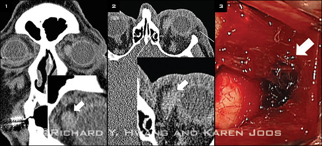
A maxillary facial CT—coronal cut (Fig. 1) and axial cut (Fig. 2)—showed expansion of the medial rectus muscle. Insets in both images show magnified views (arrows) of the medial rectus.
Because of concern about a possibly ruptured globe, the patient was taken to the operating room for globe exploration. He was confirmed to have a large conjunctival laceration and a left medial rectus hematoma with a partially lacerated medial rectus muscle (Fig. 3), but his globe was intact. The conjunctival laceration was sutured, and the injury was managed conservatively, pending resolution of the hematoma.
| BLINK SUBMISSIONS: Send us your ophthalmic image and its explanation in 150-250 words. E-mail to eyenet@aao.org, fax to 415-561-8575, or mail to EyeNet Magazine, 655 Beach Street, San Francisco, CA 94109. Please note that EyeNet reserves the right to edit Blink submissions. |