Blink
Pupillary Membrane
By Brian E. Goldhagen, MD, and Alan N. Carlson, MD, Duke Eye Center, Durham, N.C., and photographed by Tiffanie Keaton, Duke University Medical Center, Durham, N.C.
Download PDF

A 60-year-old woman underwent phacoemulsification with in-the-bag intraocular lens implantation in her left eye. She had a history of diabetes without retinopathy but no history of any inflammatory conditions; the surgery was uneventful. At the completion of the procedure, stromal hydration of the wound was not sufficient to completely seal the incision. A small air bubble was placed to help seal the internal aspect of the wound lip, and it obviated the need for a suture.
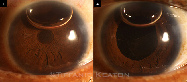
Postoperatively, pressure was normal, and the patient’s vision improved; however, in addition to the remaining air bubble, a fibrin membrane bowed forward, with pigment cells occluding the pupil (Fig. 1). Upon dilation of the patient’s pupil, the membrane stretched (Fig. 2) and separated from the pupillary border, confirming the fibrin nature of the membrane and an absence of vitreous. The patient was placed on a topical steroid four times per day and an NSAID three times per day. By her postoperative week 1 visit, the membrane was completely resolved and her uncorrected visual acuity was 20/25.
Postoperative fibrin membrane formation is thought to be caused by an immunologic response to a breakdown of the blood-aqueous barrier. It is typically seen within the first week after cataract surgery and may be associated with previous uveitis as well as systemic conditions such as diabetes mellitus.
| BLINK SUBMISSIONS: Send us your ophthalmic image and its explanation in 150-250 words. E-mail to eyenet@aao.org, fax to 415-561-8575, or mail to EyeNet Magazine, 655 Beach Street, San Francisco, CA 94109. Please note that EyeNet reserves the right to edit Blink submissions. |