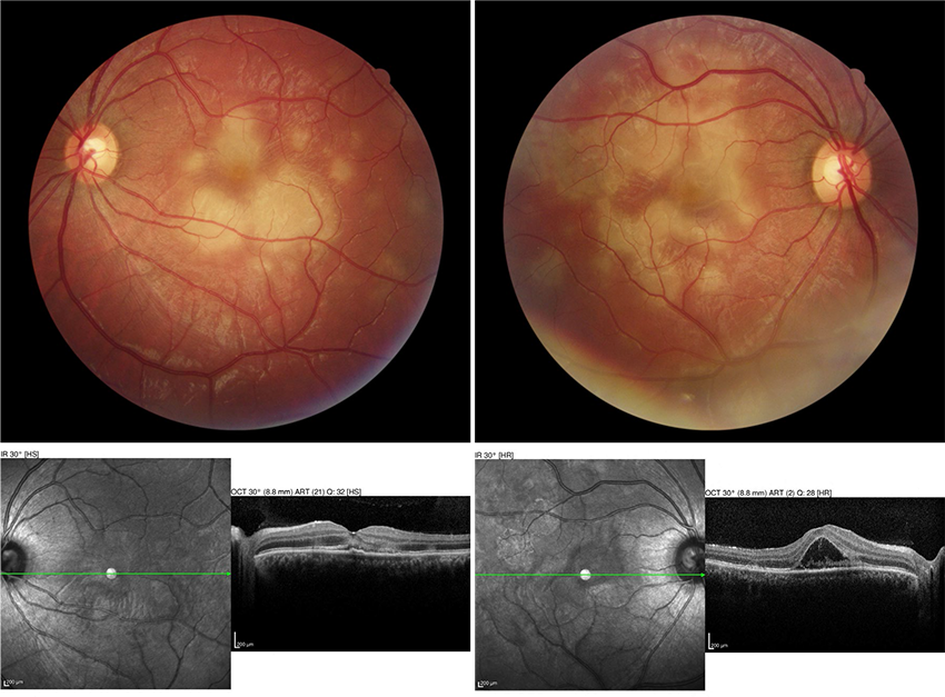
Blink
Can You Guess December's Mystery Condition?
Download PDF
Make your diagnosis in the comments, and look for the answer in next month’s Blink.

Last Month’s Blink
Apocrine Hidrocystoma
Written by Mike Rosco, MD, and Rona Z. Silkiss, MD, FACS, California Pacific Medical Center, San Francisco. Photo by Rona Z. Silkiss, MD, FACS.
A 23-year-old woman presented with a 10-month history of an enlarging mass on her left eye. Slit-lamp exam revealed a well-circumscribed 1- × 1.2-cm spherical cyst of the left superotemporal bulbar conjunctiva upon inferomedial gaze (Fig. 1). The patient requested removal of the lesion as it was irritating, cosmetically distracting, and leading to an obvious fullness of the upper eyelid (Fig. 2). Surgical excision was performed with narrow margins. Biopsy revealed an apocrine cell-lined cystic structure representing an apocrine hidrocystoma (Fig. 3).
Apocrine hidrocystomas are benign, well-defined, dome-shaped, cystic nodules with a smooth exterior. They are characterized by a double epithelial lining and an outer myoepithelial layer. Ocular apocrine hidrocystomas originate from the blocked excretion of the glands of Moll. Although usually found on the lid margin, apocrine hidrocystoma located on the conjunctiva has been documented in a few cases.
Predominantly seen in adults between the ages of 30 and 70, these formations are equally prevalent in men and women, and there is no ethnic preponderance.1
__________________________
1 Boyun K, Nam K. Int J Ophthalmol. 2012;5(2):247-248.
Read your colleagues’ discussion.
| BLINK SUBMISSIONS: Send us your ophthalmic image and its explanation in 150-250 words. E-mail to eyenet@aao.org, fax to 415-561-8575, or mail to EyeNet Magazine, 655 Beach Street, San Francisco, CA 94109. Please note that EyeNet reserves the right to edit Blink submissions. |