Blink
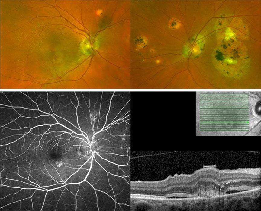
Can You Guess January's Mystery Condition?
Download PDF
Make your diagnosis in the comments, and look for the answer in next month’s Blink.

Last Month’s Blink
Contact Lens Overwear
Written and photographed by Ruben Kuruvilla, MD, Laser Eye Surgery of Erie, Erie, PA.
A 31-year-old man was referred for “epithelial edema” and linear crystal-like deposits that could not be debrided in the left cornea (Fig. 1). The patient admitted to wearing contacts for 60 consecutive days without removal and reported possible corneal abrasion of the left eye two weeks earlier when he unsuccessfully attempted to replace his monthly disposable soft lenses. At presentation, visual acuity (VA) was 20/50 in the right eye with his contact lens and 20/400 with his contact lens in the left eye, which additionally had 4+ diffuse conjunctival injection with 360 degrees of limbal blanching.
High-magnification inspection revealed a numerical dot-matrix pattern in the far inferior-temporal periphery (Fig. 2) of his left cornea consistent with a retained soft contact lens. Remarkably, the conjunctiva had grown over the top of the lens (seen as the area of limbal blanching). After blunt dissection with a Weck-Cel sponge, the edge of the lens was freed and then removed. He was started on preservative-free artificial tears and prophylactic antibiotic drops and instructed to discontinue contact lens wear. At his one-week follow-up visit, his best-corrected VA was 20/25 with no further pain, a completely clear cornea, and markedly improved conjunctival injection and chemosis of the left eye (Fig. 3).
Read your colleagues’ discussion.
| BLINK SUBMISSIONS: Send us your ophthalmic image and its explanation in 150-250 words. E-mail to eyenet@aao.org, fax to 415-561-8575, or mail to EyeNet Magazine, 655 Beach Street, San Francisco, CA 94109. Please note that EyeNet reserves the right to edit Blink submissions. |