Blink
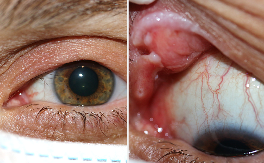
Can You Guess July's Mystery Condition?
Download PDF
Make your diagnosis in the comments, and look for the answer in next month’s Blink.

Last Month’s Blink
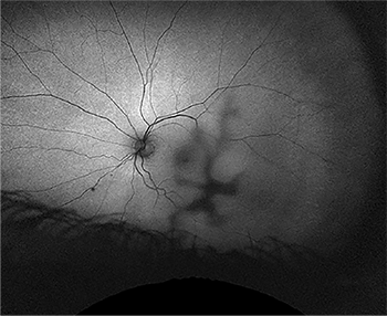
Corneal Herpetic Dendrite Identified by UWF Photography
Written by Julia Canestraro, OD, David H. Abramson, MD, FACS, and Jasmine H. Francis MD, FACS. Photo by David K. Mock, COA. All are at Memorial Sloan Kettering Cancer Center, New York. Drs. Abramson and Francis are also at Weill Cornell Medical Center, New York.

A 71-year-old man presented to the clinic with complaints of a red left eye. He was sent for imaging before seeing the physician, and this fundus autofluorescence image of his left eye was taken using ultra-widefield (UWF) fundus photography (Optos). UWF imaging is used to document and evaluate the posterior segment of the eye. However, UWF fundus imaging may also reveal anterior segment findings, which artifactually appear to be on the surface of the retina and inverted. Slit-lamp examination of the patient’s left eye confirmed the presence of a corneal herpetic dendrite. This case demonstrates that UWF fundus photography could be a useful tool for documenting potentially sight-threatening anterior segment pathology when a slit lamp is not available.
Read your colleagues’ discussion.
| BLINK SUBMISSIONS: Send us your ophthalmic image and its explanation in 150-250 words. E-mail to eyenet@aao.org, fax to 415-561-8575, or mail to EyeNet Magazine, 655 Beach Street, San Francisco, CA 94109. Please note that EyeNet reserves the right to edit Blink submissions. |