Blink
Can You Guess July's Mystery Condition?
Download PDF
Make your diagnosis in the comments, and look for the answer in next month’s Blink.

Last Month’s Blink
Alport Maculopathy
Written by Atalie C. Thompson, MD, MPH, Sharon Fekrat, MD, and Mays A. El-Dairi, MD. Photo by Michael P. Kelly, FOPS. All are at Duke Eye Center, Durham, N.C.
A 20-year-old woman with a history of sensorineural hearing loss, renal transplant for glomerulonephritis, and myopia presented for a routine eye exam. Her BCVA was 20/20 in both eyes, and her anterior segment exam was unremarkable. The fundus exam was notable for mild elevation of both optic nerve heads without blurring of the disc margins, spontaneous venous pulsations, and subtle parafoveal flecks in the macula (Fig. 1). OCT demonstrated temporal macular thinning of the inner retinal layers in both eyes (Fig. 2), as well as hyperreflective material consistent with buried optic nerve head drusen (Fig. 3).
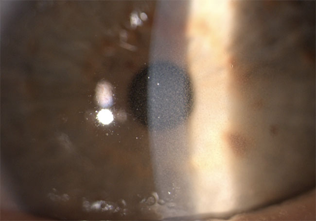
The patient’s clinical triad of glomerulonephritis, hearing loss, and ocular pathology are consistent with Alport syndrome. Ocular manifestations can include anterior lenticonus with an “oil droplet” reflex, buried optic nerve head drusen, and dot-and-fleck retinopathy with or without Bull’s-eye maculopathy. Thinning of the internal limiting membrane (ILM), retinal nerve fiber layer, retinal pigment epithelium/basement membrane, and Bruch membrane presumably stems from dysfunction in the basement membrane due to type IV collagen mutations.
While patients with temporal macular thinning on OCT may have normal visual acuity, the thinned ILM can lead to an abnormal vitreoretinal interface that may precipitate the formation of giant macular holes. Thus, patients with Alport syndrome may benefit from annual evaluation by OCT imaging for development of macular holes and buried optic nerve head drusen.
Read your colleagues’ discussion.
| BLINK SUBMISSIONS: Send us your ophthalmic image and its explanation in 150-250 words. E-mail to eyenet@aao.org, fax to 415-561-8575, or mail to EyeNet Magazine, 655 Beach Street, San Francisco, CA 94109. Please note that EyeNet reserves the right to edit Blink submissions. |