Blink
Can You Guess June's Mystery Condition?
Download PDF
Make your diagnosis in the comments, and look for the answer in next month’s Blink.

Last Month’s Blink
Dark-Without-Pressure Fundus Lesions
Written by Gaurav Gupta, MBBS, Priya Bajgai, MBBS, and Ramandeep Singh, MBBS. Photo by Arun Kapil. All are at Advanced Eye Centre, Chandigarh, India.
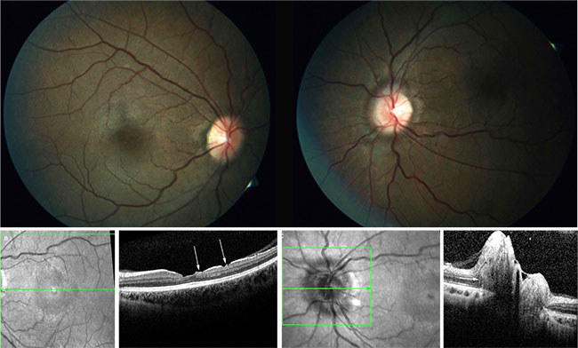
A 13-year-old boy was diagnosed with anterior uveitis in his left eye. BCVA was 20/20 in his right eye and 20/25 in his left. The fundus examination of the left eye was normal; that of the right eye showed a well-defined lesion darker than the surrounding area and sparing the macula, suggestive of a large, dark-without-pressure fundus lesion (Fig. 1, 2).1
There were no vascular abnormalities and no evidence of vitreous separation or traction. On spectral-domain OCT (Fig. 3), an abrupt transition to hyporeflectivity of the ellipsoid and outer segments of the photoreceptors was seen within the dark fundus lesion. Visual fields and full fields via electroretinogram were normal, suggesting that the lesions represent structural changes without associated functional deficits.
Dark-without-pressure lesions are asymptomatic and exhibit relative reflectivity, which occurs at the level of the outer retina.1 Specific biochemical or structural etiology remains unclear, and no functional defects have been reported in these dark areas without pressure; therefore, they do not require treatment. It has been reported1 that these lesions are transient and change shape, and they occasionally completely disappear over weeks. It has been hypothesized that they represent altered reflex at the internal limiting membrane or retinal pigment epithelium. The altered reflex has been attributed to the presence of photopigment with different density or spectral range within these lesions compared with the rest of the fundus. These dark-without-pressure focal lesions are associated with photopigment that absorbs short-wavelength light more effectively, which explains the good visibility on red-free photography.
___________________________
1 Fawzi AA et al. Retina. 2014;34(12):2376-2387.
Read your colleagues’ discussion.
| BLINK SUBMISSIONS: Send us your ophthalmic image and its explanation in 150-250 words. E-mail to eyenet@aao.org, fax to 415-561-8575, or mail to EyeNet Magazine, 655 Beach Street, San Francisco, CA 94109. Please note that EyeNet reserves the right to edit Blink submissions. |