Blink
Can You Guess March's Mystery Condition?
Download PDF
Make your diagnosis in the comments, and look for the answer in next month’s Blink.

Last Month’s Blink
Sialidosis Type 1
Written and photographed by Becky Weeks, BS, CRA, OCT-C, Moran Eye Center, Salt Lake City.
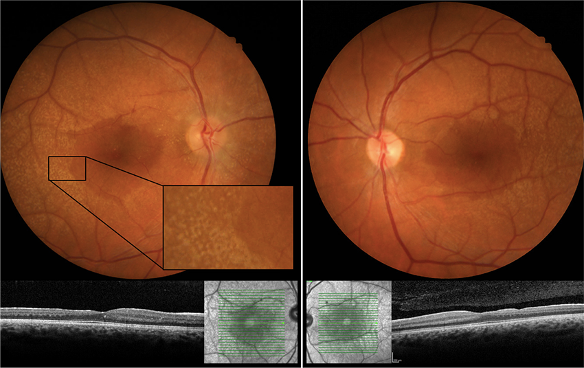
An asymptomatic 14-year-old boy was referred for a second opinion of cherry-red spots in his maculae. The best-corrected visual acuity was 20/20 in both eyes. The slit-lamp exam revealed snowflake cataracts, and the fundus exam found perifoveal graying with a cherry-red spot in both maculae. OCT showed deposits in the ganglion cell layer, and no leakage was found with fluorescein angiogram. Fundus autofluorescence (Figs. 1, 2) revealed a bull’s-eye appearance to the maculae with hypoautofluorescence surrounding a hyperautofluorescent center.
Genetic testing through Invitae Comprehensive Lysosomal Storage Disorders Panel revealed a pathogenic variant in NEU1 (neuraminidase 1) and a variant of unknown significance in SMPD1 (sphingomyelin phosphodiesterase 1).
The patient was diagnosed with sialidosis type 1. This gene mutation causes a lysosomal storage disease that is inherited as an autosomal recessive trait.
The patient’s family members came in for genetic testing and imaging. His 18-year-old brother also was diagnosed with sialidosis type 1; his father was diagnosed with MacTel (macular telangiectasia); and his mother and sister were not found to have any gene mutations.
Read your colleagues’ discussion.
| BLINK SUBMISSIONS: Send us your ophthalmic image and its explanation in 150-250 words. E-mail to eyenet@aao.org, fax to 415-561-8575, or mail to EyeNet Magazine, 655 Beach Street, San Francisco, CA 94109. Please note that EyeNet reserves the right to edit Blink submissions. |