Blink
Can You Guess May's Mystery Condition?
Download PDF
Make your diagnosis in the comments, and look for the answer in next month’s Blink.

Last Month’s Blink
Retinal Astrocytoma
Written by Catarina M. Monteiro, MD, Mário Ramalho, MD, and Inês Coutinho, MD. Photos by Pedro Lino. All are from Hospital Prof. Doutor Fernando Fonseca, Lisbon, Portugal.
A 34-year-old man reported a centrocecal scotoma in the visual field of his left eye that had been present for more than 20 years. Other than this, his ophthalmic medical history was unremarkable. The patient also revealed that he was being investigated for tuberous sclerosis by the hospital’s neurology department due to the growth of numerous benign tumors in many parts of his body, including the bladder and kidney.
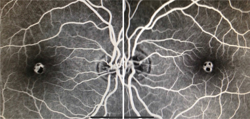
Visual acuity was 20/20 in both eyes, and anterior segment evaluation was unremarkable. Fundus examination revealed a yellowish semitransparent mulberry-like lesion, which was well circumscribed in front of the optic disc of the patient’s left eye (Fig. 1). Spectral-domain OCT (SD-OCT) of the lesion revealed moth-eaten optically empty spaces (Fig. 2). Fundus autofluorescence showed these ovoid hypolucencies as hyperautofluorescent, and the stroma of the solid lesion appeared in a pattern typical of retinal astrocytoma (Fig. 3). No other abnormal findings were evident in either eye. The patient was diagnosed with a single retinal astrocytoma, most likely associated with tuberous sclerosis. A record of the lesion was made by fundus photography (Fig. 1), and follow-up was scheduled to monitor the tumor.
Read your colleagues’ discussion.
| BLINK SUBMISSIONS: Send us your ophthalmic image and its explanation in 150-250 words. E-mail to eyenet@aao.org, fax to 415-561-8575, or mail to EyeNet Magazine, 655 Beach Street, San Francisco, CA 94109. Please note that EyeNet reserves the right to edit Blink submissions. |