Blink
Can You Guess November's Mystery Condition?
Download PDF
Make your diagnosis in the comments, and look for the answer in next month’s Blink.

Last Month’s Blink
NR2E3 Mutation
Written by Michael T. Andreoli, MD, Northwestern Medicine, Naperville, Ill. Photos by George Henry, CRA, PBT (ASCP), Wheaton Eye Clinic, Wheaton, Ill.
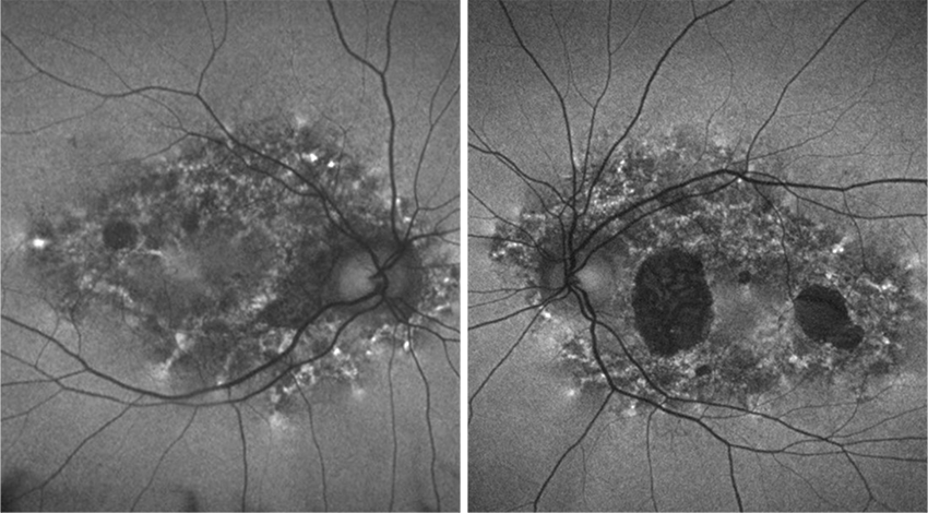
A 54-year-old man with a history of diabetes mellitus diagnosed at age 40 was referred for retinal findings on a routine exam. Visual acuity was 20/25 in each eye. Anterior segment exam revealed mild nuclear sclerosis of the lens. Dilated fundus exam demonstrated diffuse retinal pigment epithelium atrophy with pigment deposition and a bull’s-eye pattern. OCT exhibited diffuse outer retinal atrophy, spar-ing the foveal region (Fig. 1). Fundus autofluorescence showed an array of lobules and specks of hypoautofluorescence arranged in concentric rings in the macula of both eyes (as shown in Fig. 2 of the right eye).
After medication toxicities were ruled out by the patient’s history, the fundus autofluorescence findings helped to narrow down the differential diagnosis to mitochondrial conditions and certain retinitis pigmentosa variants. These unusual double concentric autofluorescence rings have been described with NR2E3 mutations. Genetic testing was performed, confirming a heterozygous variant in NR2E3, consistent with autosomal dominant retinitis pigmentosa in this patient. Notably, autosomal recessive mutations in NR2E3 have also been linked to enhanced S-cone syndrome, which tends to present with nyctalopia earlier in life.
Read your colleagues’ discussion.
| BLINK SUBMISSIONS: Send us your ophthalmic image and its explanation in 150-250 words. E-mail to eyenet@aao.org, fax to 415-561-8575, or mail to EyeNet Magazine, 655 Beach Street, San Francisco, CA 94109. Please note that EyeNet reserves the right to edit Blink submissions. |