Blink
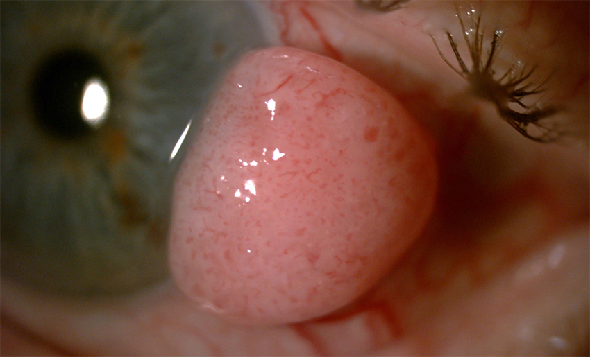
Can You Guess September's Mystery Condition?
Download PDF
Make your diagnosis in the comments, and look for the answer in next month’s Blink.

Last Month’s Blink
Bilateral Segmental Optic Disc Hypoplasia
Written and photographed by: Anamika Dwivedi, MS, Shyam Shah Medical College, Rewa, Madhya Pradesh, India.
A 19-year-old nursing student complained of occasional mild headache, which she thought might be related to refractive error. She had no significant medical or ocular history.
On examination, her uncorrected visual acuity was 20/20 in both eyes. Intraocular pressure was 12 mm Hg in both eyes, and the pupillary reactions and anterior segments were normal.
The optic discs of both eyes were smaller than normal, with cup-disc ratios of 0.5, and the cups were shifted nasally. Both optic discs showed abnormal thinning of nasal neuroretinal rim, with a large sectoral nerve fiber layer defect in nasal half of retina; this was more prominent on red free photography (Figs. 1A, 1B). OCTs revealed disc areas of 1.43 mm2 in the right eye and 1.49 mm2 in the left. OCT also showed retinal nerve fiber layer thinning (Figs. 2A, 2B). Perimetry showed a temporal hemianopia in the right eye and inferotemporal field defect in the left (Figs. 3A, 3B).
The patient was diagnosed with segmental optic disc hypoplasia. Magnetic resonance imaging of the brain, done to rule out any associated structural anomaly, was found to be normal.
Despite her optic disc hypoplasia, the patient had no visual symptoms.
Read your colleagues’ discussion.
| BLINK SUBMISSIONS: Send us your ophthalmic image and its explanation in 150-250 words. E-mail to eyenet@aao.org, fax to 415-561-8575, or mail to EyeNet Magazine, 655 Beach Street, San Francisco, CA 94109. Please note that EyeNet reserves the right to edit Blink submissions. |