Blink
Can You Guess September's Mystery Condition?
Download PDF
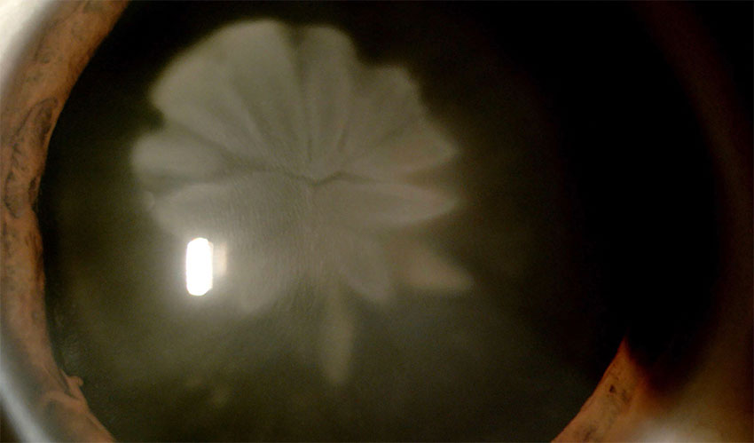
Make your diagnosis in the comments, and look for the answer in next month’s Blink.

Last Month’s Blink
Anterior Segment OCT Image of a Xen Gel Stent for Glaucoma
Written by Sylvia H. Chen, MDCM, MBA, FRCSC, Department of Ophthalmology and Visual Sciences, University of Alberta, Canada. Photos by Royal Alexandra Hospital Eye Clinic, Edmonton, Alberta, Canada.
A 73-year-old woman underwent routine and uneventful implantation of an ab interno Xen gel stent for primary open-angle glaucoma of the left eye. The next day, the 6-mm implant was noted to be in good position. The proximal end was visualized in the anterior chamber (Fig. 1A), and the distal end in the subconjunctival space, approximately 2 to 3 mm posterior to the limbus, with diffuse bleb formation (Fig. 1B). Anterior segment optical coherence tomography (AS-OCT) imaging provided similar confirmation of appropriate implant positioning (Figs. 2A and 2B; green arrow depicts the course of the cross sectional image on the right).
In this patient with blue irides, AS-OCT provided clearer visualization of the implant in the anterior chamber than did slit-lamp examination. In the early postoperative phase, when gonioscopy is often deferred to avoid putting pressure on the eye, AS-OCT can—without contact—show the position of the stent, its course through the sclera, its location under the Tenon’s tissue or conjunctiva, and the resultant bleb.
Read your colleagues’ discussion.
| BLINK SUBMISSIONS: Send us your ophthalmic image and its explanation in 150-250 words. E-mail to eyenet@aao.org, fax to 415-561-8575, or mail to EyeNet Magazine, 655 Beach Street, San Francisco, CA 94109. Please note that EyeNet reserves the right to edit Blink submissions. |