By Kavitha R. Sivaraman, MD, With Nicole R. Fram, MD, and Joshua C. Teichman, MD, MPH
Download PDF
Managing complex cataract cases can be tricky for surgeons who don’t routinely encounter corneal disease. To address this topic, Kavitha R. Sivaraman, MD, at the Cincinnati Eye Institute, convened a roundtable discussion with Nicole R. Fram, MD, at Advanced Vision Care in Los Angeles, and Joshua C. Teichman, MD, MPH, at Prism Eye Institute and the University of Toronto. In this second installment of a three-part series, these experts discuss tips for successful cataract surgery in eyes with keratoconus or a corneal graft. The final part of this series will appear in October.
Refractive Targets and Patient Counseling
Dr. Sivaraman: How does keratoconus affect your pre-op planning and IOL selection?
Dr. Fram: For patients with keratoconus, the informed consent discussion is critical. You can’t always tell which part of a keratoconic cornea the patient is looking through, and so we sometimes miss our refractive targets. I use corneal topography and wavefront analysis to evaluate the extent of higher- order aberrations. I find ray tracing aberrometers, such as the iTrace (Tracey Technologies), very helpful, and they can be used to simulate what the cornea will see clearly after surgery and the extent of the cataract’s contribution. This allows the patient to see how challenging these cases can be postoperatively. Patients with keratoconus must understand that their best vision will always be with a contact lens. However, I explain that I will do my best with lens-based surgery. These caveats are important to explain to the patient prior to surgery to set expectations and make it clear that IOL exchange may be a possibility.
Dr. Sivaraman: I always say, “The IOL we implant is a man-made lens; your natural lens can compensate for corneal aberrations in ways the artificial lens cannot. It’s possible that your vision without correction will be worse after surgery than it is now.”
Dr. Fram: I make sure I have consistent measurements on biometry, keratometry, and topography with contacts out. For a scleral lens, I ask the patient to keep the contacts out for three weeks; for a rigid gas permeable (RGP) lens, the contacts should be out for six to eight weeks if possible.
Dr. Teichman: If the patient wears an RGP or scleral lens and wants to discuss other options, you could consider offering a toric lens, but you should first ask “Are you happy with your vision when you’re wearing your glasses and your RGP lens is out?” If the answer is no, you should avoid a toric lens because these IOLs will reproduce the vision in glasses, not their vision in an RGP or scleral lens, which masks corneal aberrations. I avoid toric IOLs in patients who plan to continue wearing RGP or scleral lenses postoperatively.
Dr. Fram: I agree. Don’t push a toric lens if the patient wears hard contacts and is satisfied with them. If he or she wants a change, keep in mind that certain keratoconus cases are compatible with toric lenses, particularly eyes with a bow-tie pattern in the central 3 mm of placido imaging on corneal topography. On the other hand, patients with significant corneal aberrations need to be made aware that their best vision will be with a contact lens.
Dr. Teichman: With respect to IOL calculation, there are a number of newer formulae (e.g., Barrett True K and Kane keratoconus) that may be used, as well as methods which use K values calculated from the central zone of the cornea using devices such as the Pentacam (Oculus). Also, as keratoconus results in a hyperprolate cornea (negative spherical aberration), you may consider a neutral aspheric IOL.
 |
|
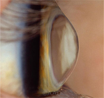
KERATOCONUS. Cataract surgery in the keratoconic eye can be complex, making the informed consent discussion indispensible.
|
Operating Through a Cone
Dr. Sivaraman: What are your practical pearls for treating these patients?
Dr. Fram: My No. 1 pearl is the steeper the cornea, the more myopic you have to aim in cataract surgery. You need to aim myopic to get to plano. This relationship was described recently in a retrospective study by Drs. Sumitra Khandelwal and Doug Koch.1 They found that hyperopic refractive prediction errors increased with steeper anterior corneas.
With a steep cone, it’s hard to determine which part of the eye is focused and which parts are out of focus. If you add room-temperature dispersive viscoelastic, such as OcuCoat (Bausch + Lomb), the imperfections in the cornea become a lot easier to operate through. An ophthalmic trypan blue solution to stain the anterior capsule also helps because these eyes have variations in surface rigidity, which can be problematic during irrigation.
Dr. Sivaraman: It’s often difficult to get corneal wounds in these eyes to hydrate and seal on their own. I have a low threshold for placing a suture in the keratome incision and sometimes even the paracentesis. I’m then careful not to remove the sutures too early—usually not until the one-month mark.
Dr. Fram: I’d add: Make your incision at the limbal vessels, take down conjunctiva, and do a bit of a scleral tunnel if necessary. Place a suture at the paracentesis site, and then avoid removing the stitches too soon.
I have placed extended depth of focus (EDOF) IOLs (e.g., Tecnis Symfony IOL [Johnson & Johnson]) in some patients with keratoconus, and there are “monofocal enhanced” IOLs that allow for better focusing power at intermediate distances. Examples are the Tecnis Eyhance (Johnson & Johnson) and Xact Mono-EDOF (Santen), which have the advantages of a wider landing zone and toric correction. Small-aperture technology also might benefit these patients. AcuFocus developed the IC-8 IOL, which is currently in the premarket approval process at the FDA.
Dr. Teichman: Although I don’t yet have personal experience with small-aperture lenses, Dr. Claudio Trindade and colleagues2 reported impressive results of a pinhole IOL implanted in patients with various types of irregular astigmatism, including keratoconus.
Cataract and Corneal Grafts
Dr. Sivaraman: For a patient who has already had a penetrating keratoplasty (PK) or a phakic Descemet membrane endothelial keratoplasty (DMEK) or Descemet stripping endothelial keratoplasty (DSEK), what goes into your pre-op evaluation?
Dr. Teichman: The patient’s vision before the cataract developed is a good indicator of visual potential. For patients who have had a PK, you want to determine what their vision was in glasses—or if they wear an RGP or scleral contact lens whether they plan to continue wearing these postoperatively. Toric IOLs are reasonable after a PK if the patient does not plan to continue wearing an RGP postoperatively and has relatively regular astigmatism centrally, but the patient must be made aware that the quality of vision will almost always be best with an RGP or scleral lens
Preoperatively, I make sure that the ocular surface is optimized because irregularities can affect the biometry, keratometry, and topography readings. I consider the indication for the graft as well. For instance, if it was indicated for herpes, the patient probably should be on a prophylactic antiviral in the perioperative period, if they are not already. I’m also careful to look for changes in the periphery of the cornea and graft because eyes with keratoconus may continue to “progress” in the periphery.
For my IOL calculations, I use the patient’s actual keratometry readings (i.e., Ks) and axial-length measurements. (This is opposed to cataract surgery in patients who may have future PK, in which case we often use K values of 45 D or audit our own results.) For the Ks, I look for consistency, and I make sure that the astigmatism aligns well with the patient’s glasses/refraction. In patients with prior PK or keratoconus, the astigmatism is almost exclusively from the abnormal cornea. Therefore, if there isn’t alignment of the refraction to auto or manual Ks, topography, and biometry, that’s a reason to avoid a toric lens.
If these measurements do align well, I think these patients can benefit from a toric lens. This is especially true nowadays because if the PK fails, we can do a DMEK or DSEK under the PK (rather than repeating the PK), so the cylinder shouldn’t change much. What Dr. Fram mentioned for keratoconus holds true for grafts as well; cover the topography, and just look at the pattern in the central 3 mm to decide whether to offer a toric lens.
I also think about how long the graft has been in place and the possibility that it might not survive the cataract surgery. Depending on the number and morphology of the endothelial cells, phacoemulsification alone is usually safe; however, there are times where cataract surgery may be combined with DMEK. With regard to spherical equivalent, if I think the graft will survive, then I aim for plano or slightly myopic. If I think the graft may not survive, I aim where I normally would for DMEK or DSEK.
Premium IOLs
Dr. Sivaraman: In a patient with previous PK who already wears an RGP contact lens, what’s your approach to selecting a premium IOL, assuming the patient is a candidate?
Dr. Fram: If the patient already wears an RGP lens or a rigid scleral lens and is happy with it, especially if there is more than 4 D of astigmatism, I tend to recommend a monofocal IOL. If the patient isn’t happy or can’t tolerate the scleral lens, and there is 4 D or less of regular astigmatism—looking at the central 3 mm—then I consider other options. As Dr. Teichman mentioned, we can now do a DMEK and preserve the graft in many cases, but there are still caveats. A very small graft might not survive a DMEK, and for a graft with a lot of volume, it can be difficult to reach the pressurization needed for retention. I try to be realistic and advise patients of all these possibilities.
EDOF and trifocal lenses are designed to be permanent, but the progression of a grafted cornea can be unpredictable. As a result, I tend to avoid those lenses in these patients. Monofocal-enhanced technology also may be an option to achieve improved depth-of-focus without loss of contrast in post-PK patients.
PK and Dense Cataract
Dr. Sivaraman: What would be your intraoperative considerations for someone with a 30- or 40-year-old PK and an endothelial cell count of 500?
Dr. Sivaraman: Dispersive viscoelastic is your friend. In these cases, I prefer a dedicated dispersive viscoelastic agent to coat the endothelium (rather than a viscoadaptive agent). I reapply it liberally, particularly when I’m operating in a shallow anterior chamber (AC) with a dense cataract.
Dr. Teichman: I use Steve Arshinoff’s soft-shell technique for viscoelastic application.3 I find that it’s gentle on the endothelium without restricting surgical maneuverability. I also avoid any incisions or manipulation at the graft-host junction.
For nucleus fragmentation, I use a chopping technique. In uncomplicated cataract surgery, I opt for a phaco hemi-flip, but I avoid this in posttransplant cataract cases because it involves slightly more energy in the AC. Instead, I chop while working as posteriorly as I safely can. Surgeons need to be mindful not just of dissipated phaco energy but also of prolonged fluid flow in the eye. If you’re spending too much time in the eye, the prolonged irrigation against the endothelium can be problematic.
Dr. Fram: I would use trypan blue for better visualization in this case. In a dense cataract, you can be deceived into thinking your visibility is good, and then all of a sudden, you can’t identify the AC and the red reflex is insufficient.
Post-Op Edema
Dr. Sivaraman: Patients with corneal grafts—especially those with older grafts and low endothelial cell count— tend to have edema after cataract surgery. How do you address this?
Dr. Fram: To reduce inflammation and edema, you can give a subconjunctival injection of triamcinolone acetonide intraoperatively.
Dr. Sivaraman: Many post-PK patients are already on long-term steroid treatment to prevent rejection. For uncomplicated eyes, I usually taper steroids after cataract surgery over four weeks. However, in most postgraft patients, I tend to taper the steroid very slowly after cataract surgery and, of course, I never go lower than the maintenance dosage.
___________________________
More at the meeting. Don’t miss the 20th Spotlight on Cataract session at AAO 2021. The morning’s program will be capped off with the Charles D. Kelman Lecture, titled “Niche Devices for Special Eyes,” presented by Michael E. Snyder, MD, of the Cincinnati Eye Institute. When: Monday, Nov. 15, from 8:00 a.m. to noon. Where: The Great Hall, Ernest N. Morial Convention Center, New Orleans.
___________________________
1 Khandelwal S et al. Predictive accuracy of intraocular lens calculation formulas in eyes with keratoconus. Presented at: ASCRS; May 3-7, 2019; San Diego.
2 Trindade CC et al. J Cataract Refract Surg. 2017;43(10):1297-1306.
3 Arshinoff SA. J Cataract Refract Surg. 1999;25(2):167-173.
___________________________
Dr. Fram is managing partner at Advanced Vision Care in Los Angeles. Relevant financial disclosures: Johnson and Johnson Vision: C.
Dr. Sivaraman is a cornea and cataract surgeon at the Cincinnati Eye Institute in Cincinnati. Relevant financial disclosures: None.
Dr. Teichman is a cornea and cataract surgeon at Prism Eye Institute and the University of Toronto, in Toronto, Ontario, Canada. Relevant financial disclosures: Alcon: C; Bausch + Lomb: S.
For full disclosures and the disclosure key, see below.
Full Financial Disclosures
Dr. Fram Alcon: C,L; Allergan: C; Beaver-Visitec International: L; CorneaGen: C,O,L; Dompé: C,L; Johnson & Johnson Vision: C; MicroSurgical Technology: L; Novartis: L; Ocular Science: O; Ocular Therapeutix: C,L.; CorneaGen: C,S.
Dr. Sivaraman None.
Dr. Teichman Aequus: C; Alcon.: C; Allergan: C; Bausch + Lomb: S; Labtician Ophthalmics: C; Novartis: C; Santen: C; Shire: C; Sun: C.
Disclosure Category
|
Code
|
Description
|
| Consultant/Advisor |
C |
Consultant fee, paid advisory boards, or fees for attending a meeting. |
| Employee |
E |
Employed by a commercial company. |
| Speakers bureau |
L |
Lecture fees or honoraria, travel fees or reimbursements when speaking at the invitation of a commercial company. |
| Equity owner |
O |
Equity ownership/stock options in publicly or privately traded firms, excluding mutual funds. |
| Patents/Royalty |
P |
Patents and/or royalties for intellectual property. |
| Grant support |
S |
Grant support or other financial support to the investigator from all sources, including research support from government agencies (e.g., NIH), foundations, device manufacturers, and/or pharmaceutical companies. |
|