Blink
Choroidal Tuberculoma
By Inês Coutinho, MD, Cristina Santos, MD, and Manuela Bernardo, MD, Hospital Prof. Doutor Fernando Fonseca, Amadora, Portugal
Photo by Susana Teixeira, Hospital Prof. Fernando Da Fonseca E.P.E., Lisbon, Portugal
Download PDF

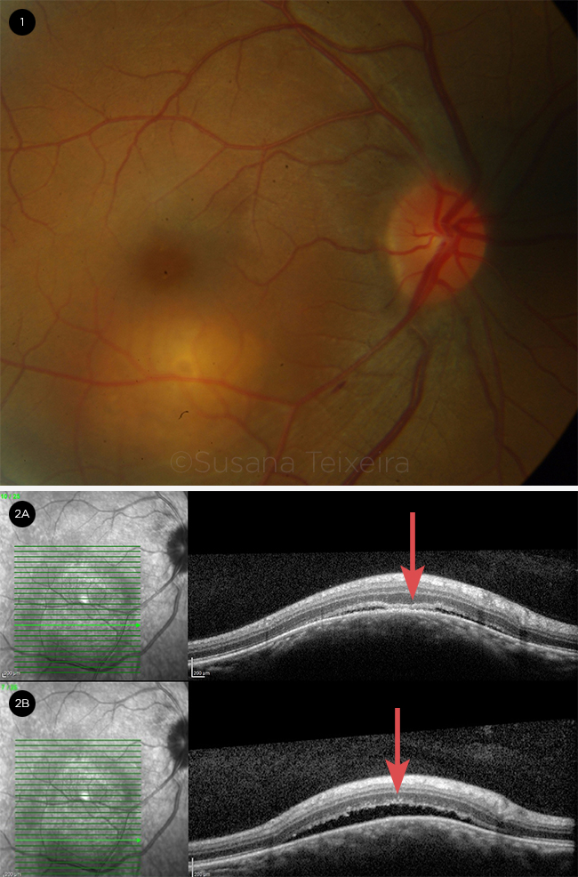
A 39-year-old African woman, 30 weeks pregnant, presented in the emergency room with blurred vision in her right eye. Ocular examination revealed a well-defined yellow subretinal mass of about 2 disc diameters, located near the inferotemporal arcade in the right eye (Fig. 1). Optical coherence tomography (Fig. 2) showed serous detachment of the neurosensory retina (Fig. 2B) and an area of localized adhesion between the neurosensory retina and retinal pigment epithelial–choriocapillaris layer (contact sign, Fig. 2A). The differential diagnosis included ocular tuberculosis, sarcoidosis, choroidal amelanotic melanoma, and metastatic choroidal tumor.
After 2 weeks, the patient developed dyspnea, fatigue, and fever. Systemic evaluation established the diagnosis of miliary tuberculosis, and a presumptive diagnosis of ocular tuberculosis (TB) with choroidal tuberculoma was made. Antibiotics were started, but she died from septic shock shortly after giving birth to a baby at 33 weeks of gestational age and low birth weight. After delivery, the baby began treatment, but TB tests (Mantoux and interferon gamma release assay) came out negative twice.
| BLINK SUBMISSIONS: Send us your ophthalmic image and its explanation in 150-250 words. E-mail to eyenet@aao.org, fax to 415-561-8575, or mail to EyeNet Magazine, 655 Beach Street, San Francisco, CA 94109. Please note that EyeNet reserves the right to edit Blink submissions. |