Download PDF
Periodically, the evening news brings the most unwelcome of images: graven-faced but determined doctors treating casualties from the latest mass disaster—earthquake or tornado devastation, airshow or racetrack collisions, oil-rig or coal-mine explosions, or, perhaps most troubling, the rampages of domestic terrorists or deranged gunmen.
One such physician in the news was Lynn Polonski, MD, who appeared on televisions across the country in January 2011, describing the injuries sustained by the now-retired Arizona congresswoman Gabrielle Giffords in a shooting that killed six and injured 12 of her constituents. “I received a frantic call from our chief of trauma, who didn’t have time to describe what had happened or to whom. He just shouted ‘Get your [bleep] in right now!’ I soon discovered that Rep. Gabby Giffords was the patient presenting—and presenting with severe neurologic and ophthalmic trauma.” Dr. Polonski is a clinical assistant professor of ophthalmology at the University of Arizona in Tucson, who also maintains a private oculoplastic and comprehensive eye practice there.
Vikram D. Durairaj, MD, is another surgeon familiar with the tragedy and publicity of mass murder. A professor of ophthalmology and otolaryngology and the chief of oculoplastics and orbital surgery at the University of Colorado Hospital (UCH) in Aurora. Dr. Durairaj was covering orbital trauma at UCH the night of the Aurora Theater shootings last July. Twelve people were killed, and many of the 58 wounded were taken to UCH for medical attention.
Unexpected gun violence on this scale is reminiscent of medical service in a military setting. And, in fact, civilian ophthalmologists can benefit greatly from the experience gained by their counterparts in war zones on such critical points as establishing treatment priorities and planning for future oculofacial repair. Although the focus of this story is on injuries produced by firearms, these principles apply to any type of major oculofacial trauma.
|
Bone Repair
|
 |
|
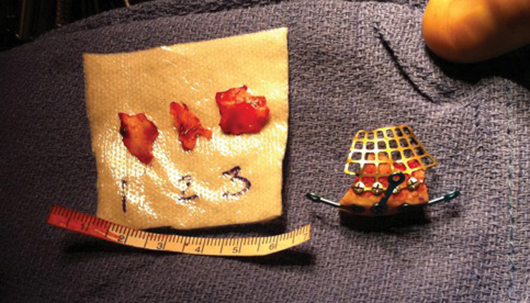
(Left) Bony fragments recovered from the orbital apex of a gunshot victim; (right) superior orbital rim and roof with titanium plates for reconstruction.
|
On the Home Front: Gunshot Wounds in the ER
Scope of the problem. The CDC reports that, in 2010, firearms were involved in 19,038 suicides, 11,015 homicides, and 31,513 injuries in the United States.1 According to a report by the University of Pennsylvania, “Firearm injury in the United States averaged 32,538 deaths annually between 1970 and 2002. It is the second leading cause of death from injury after motor vehicle crashes and, in several states, is the leading cause of injury death.”2 Given such statistics, any ophthalmologist who takes emergency calls may face the necessity of managing gunshot wounds.
Trauma’s trajectory. Beyond the sheer force and velocity produced by firearms, Dr. Polonski said that each incident of gun trauma is unique in its details, depending on:
- The caliber and velocity of the projectile
- The proximity of the firearm to the target
- The projectile’s entry and exit trajectory
- Foreign debris in the wound
Dr. Durairaj agreed with those variables and added some nuances:
- The difference in kinetic energy between handguns and automatic or assault-type weapons
- The wide spectrum of ophthalmic injury (e.g., corneal penetration or perforation, globe rupture, and orbital fracture)
- The probability that the injury is not confined to the eye and is a multisystem trauma
The last point means that the ophthalmologist will most likely be functioning as part of a team, whose chief priorities will not necessarily include vision.
Critical Considerations
Dr. Polonski said that the priorities trauma surgeons observe in an ER are uniform and well established, replicating those followed by military physicians who treat victims of combat trauma: Address threats to the patient’s life, then to vision, then to other organs and limbs.
Keep them breathing, stop the bleeding. Dr. Durairaj agreed, saying that the underlying principles that guided him during the night of the theater shootings are the same that apply in a combat zone. This algorithm is relevant not only to gunshot wounds but also to any traumatic injury:
- Clear the patient’s airway and ensure that unobstructed breathing is maintained
- Control hemorrhaging and blood pressure
- Limit insults to the brain and nervous system
He added that in most ER situations, the patient will be stabilized by other physicians—trauma surgeons for shots to the torso, neurosurgeons in the case of intracranial trauma—before the ophthalmologist can step in. Deferring to more urgent lifesaving measures may cause unavoidable delays in dealing with ophthalmic crises, Dr. Durairaj said.
Assess the possibilities. In some cases, however, emergency ophthalmic surgery may need to be performed before or in conjunction with other critical procedures. In recounting the Aurora shootings, Dr. Durairaj said, “I arrived and encountered a patient who had sustained a close-range, devastating gun blast to the face. A projectile had been fired through his eye and orbit and was lodged in his brain. The globe was devastated, leaving no possibility for salvage whatsoever.”
Primary enucleations are exceptionally rare in civilian ERs, he noted, and in more forgiving circumstances, he would calmly discuss each situation’s possibilities—or inevitabilities—with the patient. “But in this tragic case, we knew our patient would not be conscious for some time, and all surgeons present concurred there was no chance of even piecemealing the globe together. So we surgically removed the eye’s remnants, after which the neurosurgeons decompressed his intracranial pressure and proceeded with primary repairs to his brain.”
Ocular Priorities: What Can’t Wait
Protect the globe first. Well-versed in the hazards of treatment delays is Col. (Ret.) Robert A. Mazzoli, MD, former Consultant to the Surgeon General of the U.S. Army and former chief of ophthalmology at Madigan Army Medical Center in Tacoma, Wash. “Study after study has shown that the longer the delays of primary globe repair, the more likely the patient will lose vision and the higher the risk of endophthalmitis. In severe trauma, a normal IOP of 10 to 20 can drop to zero, and hypotony then causes choroidal expulsion. In the combat theater, just getting ocular contents back into the globe and the globe into the orbit is incredibly urgent; and in those initial moments, exact anatomic precision is not as important as getting the eye initially closed, with a more meticulous closure to follow quickly. I would say the window of vitality is under 24 hours for globe and lid repair, but orbit repair can wait longer, and we even tend to wait just to allow the periocular swelling to go down.”
The importance of lowering IOP. Dr. Polonski agreed that orbital trauma can wait longer than globe repairs, but he is emphatic about relieving any elevated intraocular pressure if there is compression on the globe. “High pressures can injure an optic nerve that was previously unscathed by the initial trauma and can cause central retinal artery and vein occlusions. An otherwise visually intact eye in this situation can lose visual function completely. So if you find elevated IOP, you will never go wrong by doing a canthotomy, which allows orbital tissues compressing the optic nerve to relax. Protocols [used] for teaching ER docs, especially in rural areas, offer this as a first line of defense if no ophthalmologist is available.” In selected cases, he also administers IOP-lowering drugs; his choices in trauma cases are timolol 0.5 percent and brimonidine, which may also provide neuroprotective properties.
If IOP is in the 40s or more, and pressure is not controlled despite these drugs, canthotomy, and cantholysis, consider adding topical dorzolamide or oral acetazolamide, he said.
An Approach to Primary Repair
When the way is clear to initiate primary repairs of eye trauma, enucleation is sometimes the only resort. But when the globe is largely intact, Dr. Polonski can predict some of the more typical wounds and approaches. “For projectiles that traverse the temple, some frontal parietal bone is usually blown off, and shock waves tend to collapse the orbital roof and proptose the globe, leaving metal fragments all along the course of the trajectory. It can also leave a dural tear, allowing brain matter to herniate into the orbit. In that case we immediately perform a craniectomy and stabilize the bleeding. We also dilate the patient and assess the optic nerves. If IOP is high, we decompress the patient with a lateral canthotomy and inferior and superior cantholysis.”
Dr. Polonski added that the craniectomy and canthotomy, together and separately, allow the orbital tissues and bony fragments that may be pressing anteriorly on the optic nerve and, posteriorly, on the globe, to relax. If he wishes to further reduce pressure, he may administer drugs (see “The Importance of Lowering IOP,” above).
“Although repairs in an emergency setting cannot be expected to restore pretrauma anatomy, we then repair the roof of the orbit by making a brow incision, performing a frontal craniotomy, and removing the superior orbital rim, along with any remaining bullet and bony fragments in the apex of the orbit. We also remove any remaining devitalized brain tissue and patch the dural tears. Finally, we replace the missing roof components with a titanium plate. The plate, perhaps 0.4 mm thick, is screwed down onto the roof of the orbit, close to the superior orbital rim, so it has good rigidity.”
Amazingly, in spite of the insults to the orbit, muscles, globe, cranium, and brain, some patients may not lose substantial visual function, Dr. Polonski said. “If cranial nerve V1 is not affected and the patient is seeing well, they might achieve functional vision with only a slight field defect due to traumatic optic neuropathy. With significant trauma you may have some neuronal injury to rectus muscles, muscle atrophy, or loss of skin and volume, but you do your best to make the patient happy, stressing both function and cosmesis.”
Picking Your Battles
“You have to pick your battles in trauma,” said Dr. Polonski. “Preserving the vitality of the globe and optic disc comes first, but you can’t then ignore the lids or orbit.”
Dr. Mazzoli agreed. “If you don’t have a good lid protecting the globe, you can actually lose the eye, or if you have disorganized contents, you can lose the eye. In one case I know of, the eyes were horribly, devastatingly injured, and the doc would have been justified to primarily enucleate both. Stateside, the patient would be facing total blindness. Ironically, given the terror and panic of combat, but also logically, thanks to the honor and motivation of service, military surgeons tend to pull out all the stops. This patient went from NLP in both eyes to some vision in one eye because of the tenacity of the ophthalmologist who first saw the patient and of the follow-on surgeons stateside.
“There are no hard rules, sometimes, especially when the globe and orbit are both decimated. There are cases where the socket is so bad that it has to be addressed before the globe. The globe is what determines vision, of course, but not addressing orbital trauma might leave the threat to vision intact,” he said.
The bottom line, said Dr. Durairaj, is endowing critical surgery with the same goals as planned surgery. “Just as for the most routine, elective procedures, emergency, survival-critical surgeries demand that we confirm anatomy, that we think about safety landmarks, work on surgical planes that respect safe dissection principles, and account for efficient exposure and hemostasis.”
___________________________
1 Centers for Disease Control and Prevention. National Vital Statistics Reports. Deaths: Preliminary Data for 2010. January 2, 2012. www.cdc.gov/nchs/data/nvsr/nvsr60 /nvsr60_04.pdf. Accessed Nov. 5, 2012.
2 Firearm Injury in the U.S. Firearm & Injury Center at Penn. www.uphs.upenn.edu/ ficap/resourcebook/pdf/monograph.pdf. Accessed Nov. 5, 2012.
___________________________
Drs. Durairaj, Mazzoli, and Polonski report no related financial interests.
___________________________
NOTE: The views expressed in this article are those of the author(s) and do not necessarily reflect the official policy or position of the U.S. Departments of the Navy, Army, or Air Force; the U.S. Department of Defense; or the U.S. government.