Download PDF
With more than a million retinal ganglion cells (RGCs) in the human eye, all with vast thickets of dendrites, it stands to reason that evolution must have developed ways to organize the electrical signals that these cells gather for sending to the optic nerve. But what are the structural features that enable coordination of all this activity?
Researchers at Duke University in Durham, North Carolina, have published two papers that they believe help an-swer that question.1,2 The papers describe the relative arrangements of retinal mosaics, collections of cells that divide up visual space into small regions called receptive fields, one per ganglion cell.
 |
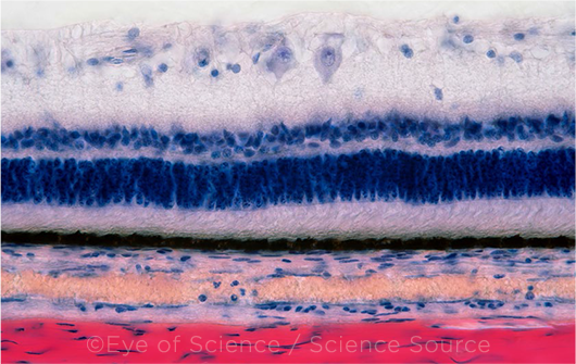
RETINAL NUANCES. Different sets of retinal neurons are sensitive to individual stimuli.
|
Stacked mosaics. There are roughly 40 types of such mosaics, encoding visual features from motion to contrast, but until recently, it was unclear how the mosaics were arranged.
Last April, the Duke researchers reported that some of these mosaics were carefully positioned with respect to one another.1 In the group’s second paper,2 they used computational modeling to compare their laboratory observations to the optimized mosaic layouts predicted by an idea called “efficient coding theory.” The real-world data and the model’s predictions matched.
“The retina is not one mosaic. It’s a whole bunch of stacked mosaics. And each of these mosaics encodes something different about the visual field,” said coauthor Greg D. Field, PhD. “The depth that the dendrites reach in the retina is kind of an addressing scheme.” That is, when the dendrites reach a deeper position, they receive one type of information—and if they are in a shallower position, they receive another.
Subtle organization scheme. The modeling studies also showed that the processing efficiency of the mosaics was affected by the pattern in which they were laid out, said coauthor John Pearson, PhD. “There’s a level of organization to the retina that is even greater than what we had thought was there. The very intricate positioning of the mosaics with respect to one another tells us that their various alignments are probably more important [than we realized].”
—Linda Roach
___________________________
1 Roy S et al. Nature. 2021;592(7854):409-413.
2 Jun NY et al. Proc Natl Acad Sci. 2021;118(39):e2105115118.
___________________________
Relevant financial disclosures: Drs. Field and Pearson—None.
For full disclosures and the disclosure key, see below.
Full Financial Disclosures
Dr. Ambati Allergan: C; Biogen: C; Boehringer-Ingelheim: C; DiceRx: O; iVeena Delivery Systems: O; iVeena Holdings: O; Inflammasome Therapeutics: O; Immunovant: C; Janssen: C; Olix Pharmaceuticals: C; Retinal Solutions: C; Saksin LifeSciences: C; University of Kentucky: P; University of Virginia: P.
Dr. Field None.
Dr. Pearson None.
Dr. Sallam None.
Disclosure Category
|
Code
|
Description
|
| Consultant/Advisor |
C |
Consultant fee, paid advisory boards, or fees for attending a meeting. |
| Employee |
E |
Employed by a commercial company. |
| Speakers bureau |
L |
Lecture fees or honoraria, travel fees or reimbursements when speaking at the invitation of a commercial company. |
| Equity owner |
O |
Equity ownership/stock options in publicly or privately traded firms, excluding mutual funds. |
| Patents/Royalty |
P |
Patents and/or royalties for intellectual property. |
| Grant support |
S |
Grant support or other financial support to the investigator from all sources, including research support from government agencies (e.g., NIH), foundations, device manufacturers, and/or pharmaceutical companies. |
|
More from this month’s News in Review