By J. Javier Servat, MD, Evan H. Black, MD, Aleksey Mishulin, BS, and Geoffrey J. Gladstone, MD; Edited by Ingrid U. Scott, MD, MPH, and Sharon Fekrat, MD
This article is from August 2012 and may contain outdated material.
When considering blepharoplasty as a primary procedure, a surgeon must keep in mind the important relationships among the forehead, eyebrows, and upper eyelids. Many times, patients are referred for “crowded” upper eyelids. Often, the culprit is actually the eyebrow. This may be corrected with forehead surgery instead of, or in conjunction with, eyelid surgery.
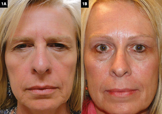
Endoscopic forehead lift can provide eyebrow lift while minimizing postoperative scarring and tissue disruption. The goal is not only to elevate the brow but also to decrease forehead rhytides (wrinkles or skin folds), decrease vertical glabellar rhytides, improve lateral canthal hooding and rhytides, and decrease the infrabrow skin area (Figs. 1A, 1B).
 |
|
MULTIPLE ACTIONS. The endoscopic forehead lift affects more than just the position of the eyebrows, as shown in this set of before (1A) and after (1B) images. The patient also underwent bilateral upper eyelid blepharoplasty.
|
Clinical Evaluation
Brow ptosis should be suspected in any patient who has redundant upper eyelid skin, even if the eyebrow appears to be in a normal position. Patients often manually lift their eyebrow when they want eyelid surgery, an action known as Flower’s sign. This is a cue to the surgeon to evaluate the patient for brow and forehead surgery in addition to, or instead of, eyelid surgery.
During the examination, the patient should be given a handheld mirror. The mirror allows patients to describe their goals. It will also give the surgeon an opportunity to point out the correct position of the brow and the possible contribution of eyelid skin and blepharoptosis, as well as to outline realistic surgical expectations. To measure the true level of the brow, have patients gently close their eyes while the surgeon’s hand is placed above the brow to fixate the forehead position in the relaxed state, and then have them gently open their eyes.
It is important to note that patients may have attempted to alter their brow position cosmetically. Patients may epilate their lateral or inferior brow cilia to create an illusion of a normal brow position or attempt to correct a low brow appearance by tattooing the eyebrows. The permanent nature and frequent nonanatomical position of eyebrow tattoos present unique challenges and should be discussed with these patients before surgery.
Patients should be warned preoperatively to expect cranial nerve V1 hypoesthesia, transient facial neuropraxia, and possible alopecia near the incision sites lasting up to three months. Preoperative discussion about the kind of fixation device is also important, as the patient may discover the anchors on palpation.
Surgical Considerations
The position of the eyebrow, and consequently the brow ptosis, is affected by brow elevator and depressor muscles, genetics, gravity, and the patient’s natural expression. Although each eyebrow has its own shape, position, and contour, the female eyebrow should lie approximately at or above the superior
orbital rim. The brow should have an arched shape, with the tail of the brow
positioned higher than the medial aspect. The peak of the arch should be two-thirds of the distance of the brow from the medial aspect. In contrast, the male eyebrow should be at the level of the superior orbital rim with a flatter, more horizontal configuration.
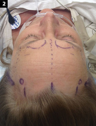
When planning brow surgery, the surgeon must consider the function and corresponding physiologic rhytides of the frontalis, orbicularis oculi, depressor supercilii, corrugators, and procerus muscles. Planning is important to avoid damaging the frontal branch of the facial nerve, the supraorbital nerves, and the supratrochlear nerves, as injury can result in paralysis and hypoesthesia (Fig. 2).
 |
|
PLANNING. Preoperative marking should include notations to protect nerves during later dissection, as injury can result in paralysis and hypoesthesia of the forehead and scalp.
|
Surgical Technique
Marking. The right and left temporal crescents, denoting the fusion of periosteum, deep temporal fascia, and temporoparietal fascia at the conjoined tendon, are palpated and marked. Marks are then made at the right and left lateral orbital rims at the level of the lateral canthal angle. These marks denote the area of the zygomaticotemporal (sentinel) vein and should be the lateral limit of the dissection.
The supraorbital notch is then palpated on each side, and a “safety zone” of approximately 2 cm is drawn around these notches to protect the supraorbital and supratrochlear nerves during later dissection. One central vertical and two paramedian vertical incision sites are then marked just posterior to the hairline. The central incision is marked at or 1 cm behind the hairline, extending 1.0 to 1.5 cm in length. The paramedian incisions are marked approximately 4.5 cm lateral to the central incision site. Alternatively, in males, a single central elliptical incision can be placed in a coronal orientation approximately 1 cm behind the hairline. Bilateral simple or elliptical incisions are then marked in the temporal areas.
The hair should not be shaved, but it can be twisted with rubber bands to maximize visualization of the surface during surgery.
Anesthesia. Endoscopic forehead lifting can be performed under local anesthesia with sedation or under general anesthesia. A bilateral supraorbital nerve block is obtained using a 50/50 mixture of 2 percent lidocaine with 1:100,000 epinephrine and 0.75 percent bupivacaine with 1:100,000 epinephrine. Additional local anesthesia using the same mixture is then administered along the surgical markings, across the entire forehead.
Incision and dissection. A scalpel is used to incise the temporal skin over the elliptical marking on each side. Further dissection exposes the deep temporal fascia. The plane of dissection should be between the superficial and the deep temporal fascia. The endoscope is introduced through the temporal incision above the deep temporal fascia. Continuing in each temporal pocket, dissection is carried out toward the lateral canthal angle. The lateral canthal ligament is then detached under endoscopic visualization.
The surgeon then makes the central and paramedian incisions, using a blade to incise the skin and scalp down to the periosteum. A periosteal elevator is then used to blindly release the periosteum across the forehead.
Under direct visualization, the supraorbital nerve is located and avoided. A supraperiosteal pocket is then formed above the bridge of the nose to address the depressor muscles. Using blunt dissection, the tissues are moved side to side to separate the muscles for better visualization. The corrugators are avulsed rather than cut. The procerus and depressor supercilii muscles are avulsed in a similar fashion. Using blunt and sharp dissection, the temporal crescents are released on each side, joining the subperiosteal and deep temporal fascial planes.
Fixation. Following adequate release of the periosteum, the surgeon then performs the fixation. Many methods are available to fixate the scalp, including the Endotine (Coapt Systems) and the Ultratine (Coapt Systems), both of which use biodegradable implants that attach to the skull. The superficial temporal fascia is closed with deep, buried absorbable or nylon sutures, and the skin is closed with surgical staples.
Postoperative Considerations
A firm dressing is placed around the forehead to reduce postoperative swelling and is left in place for 24 to 48 hours. Antibiotics and analgesic and anti-inflammatory medication may be provided at the surgeon’s discretion. Patients can usually return to work in three to five days, and the staples are removed in seven to 10 days.
Summary
This procedure is effective and long lasting and is associated with a shorter healing time and less scarring than traditional brow-lifting methods.
The key to success is meticulous release of the periosteum and brow depressor muscles. Careful dissection deeper than the superficial temporal fascia and avoidance of the frontal branch of the facial nerve, the supraorbital nerves, and the supratrochlear nerves are crucial.
___________________________
Dr. Servat is assistant professor of ophthalmology at Yale University. Drs. Black and Gladstone are associate professors of ophthalmology, and Mr. Mishulin is a medical student; all three are at Wayne State University in Detroit. The authors report no related financial interests.