Download PDF
Contrary to long-standing ophthalmic dogma, immune privilege in the crystalline lens does not exist, scientists investigating the intraocular response to eye injury have discovered. Instead, the lens and associated structures should be regarded as immune-quiescent.1
“While the lens is avascular, it’s not an immune-privileged tissue, and this is a huge sea change in the way we think about things. Everyone, including myself, just presumed that because this tissue was avascular there would be no source of immune cells” to protect the lens, said senior author A. Sue Menko, PhD, at Thomas Jefferson University in Philadelphia.
 |
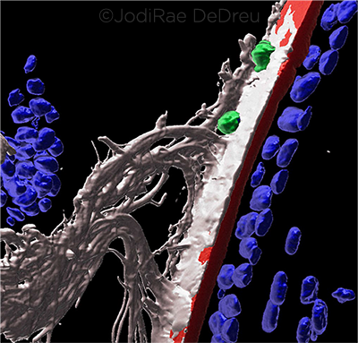
RESPONSE. At day 1 following corneal wounding, 3D surface structure imaging shows immune cells (CD45+, green) migrating within ciliary fibrils (MAGP1+, white) that extend along the surface of the matrix capsule that surrounds the lens (perlecan+, red). The ciliary zonules (white) are also evident, as are the nuclei (blue).
|
A surveillance response to corneal injury? In their experiments in mice, Dr. Menko and her colleagues found that the ciliary zonules, which contain a reservoir of two likely immune-mediator molecules, MAGP1 and TSP-1, react to corneal injury by recruiting leukocytes to the lens. Scanning electron microscope images showed that the immune cells travel along the zonular fibers, but they can also migrate onto the capsule and sometimes into the lens itself.
“We imagine what we’re describing is a protective response to the lens, as the cornea is getting repaired. It’s not an overabundance of immune cells. It looks like a surveillance response,” Dr. Menko said.
Robustness of immune response. Patient-specific cofactors, such as genetics and concurrent ocular inflammation, appear to influence the robustness of the immune response and its potential to be pathologic, she said.
However, research has shown that some of the recruited immune cells acquire a myofibroblast phenotype and begin producing a fibrotic collagen matrix. Fibrosis triggered by these cells might explain the genesis not only of posterior capsular opacification after cataract surgery but also of anterior subcapsular cataracts associated with corneal wounds, Dr. Menko said. She and coauthor Mary Ann Stepp, PhD, at George Washington University in Washington, D.C., along with Rachel R. Caspi, PhD, of the NEI, are investigating this possibility by studying the movement of autoimmune cells into the lens and resulting cataract formation in patients with inflammatory conditions such as uveitis. They hope to publish their results within the next year, she said.
Obvious in hindsight? The researchers’ paradigm-shifting conclusion that the lens does not have immune privilege might seem surprising at first—but it may appear less startling in retrospect, Dr. Menko said. “In science, sometimes we believe things because they’re dogma, but if you think about it you realize that those things don’t make sense,” she said.
“We began looking for signs of an immune response to the lens because it just seemed against all logic that you’d have a tissue that is so crucial throughout a lifetime, but which evolved in such a way that the body would not try to protect or repair it.”
—Linda Roach
___________________________
1 DeDreu J et al. FASEB J. Published online May 25, 2020.
___________________________
Relevant financial disclosures—Dr. Menko: None.
For full disclosures and the disclosure key, see below.
Full Financial Disclosures
Dr. Mainster Ocular Instruments: C.
Dr. Menko None.
Dr. Niffenegger None.
Dr. Tonorezos This study was supported by the National Cancer Institute’s Intramural Research Program, a Sloan-Kettering Institute of Cancer Research Cancer Center Support Grant (P30CA008748), and the Meg Berté Owen Fund. The funding sources had no role in the study design, data acquisition, analysis, manuscript preparation, or decision to submit this work.
Disclosure Category
|
Code
|
Description
|
| Consultant/Advisor |
C |
Consultant fee, paid advisory boards, or fees for attending a meeting. |
| Employee |
E |
Employed by a commercial company. |
| Speakers bureau |
L |
Lecture fees or honoraria, travel fees or reimbursements when speaking at the invitation of a commercial company. |
| Equity owner |
O |
Equity ownership/stock options in publicly or privately traded firms, excluding mutual funds. |
| Patents/Royalty |
P |
Patents and/or royalties for intellectual property. |
| Grant support |
S |
Grant support or other financial support to the investigator from all sources, including research support from government agencies (e.g., NIH), foundations, device manufacturers, and/or pharmaceutical companies. |
|
More from this month’s News in Review