Blink
Intraorbital Foreign Body Accompanied by Sclopetaria
By Sanket U. Shah, MD, and F. Ryan Prall, MD, and photographed by Tom Egnatz, CRA, Eugene and Marilyn Glick Eye Institute, Indiana University School of Medicine, Indianapolis
Download PDF

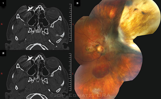
A 12-year-old otherwise healthy boy presented following a BB pellet injury to the right eye, which had visual acuity of 20/400 and a relative afferent pupillary defect. Maxillofacial computed tomography (CT) showed the pellet lodged near the medial rectus insertion (Figs. 1, 2).
Although there was no globe rupture, the patient sustained a superonasal traumatic chorioretinal rupture (sclopetaria), a rhegmatogenous retinal detachment, retinal hemorrhages, and commotio retinae. Following surgical removal of the pellet and repair of the retinal detachment with scleral buckle and laser retinopexy, we noted extensive macular pigment changes, resolving hemorrhage, and fibrotic scar at the sclopetaria site superonasally (Fig. 3). Final visual acuity was counting fingers.
Sclopetaria is a full-thickness break of retina and choroid due to a high-velocity object striking or passing close to, but not penetrating, the globe. Rapid deformation causes the chorioretinal unit to split and retract, exposing the sclera. Most cases are managed by observation and have a poor visual outcome. The bright appearance of the BB pellet is an artifact resulting from entrapped air and soft tissue near the pellet.
| BLINK SUBMISSIONS: Send us your ophthalmic image and its explanation in 150-250 words. E-mail to eyenet@aao.org, fax to 415-561-8575, or mail to EyeNet Magazine, 655 Beach Street, San Francisco, CA 94109. Please note that EyeNet reserves the right to edit Blink submissions. |