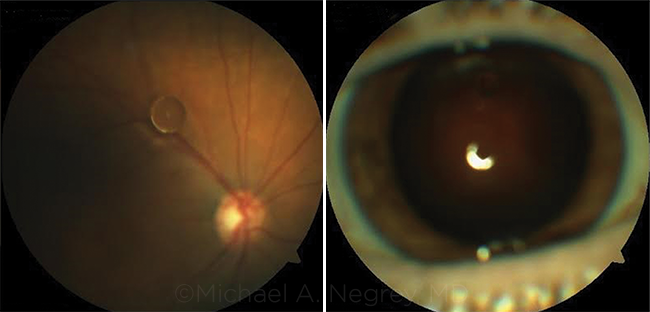
Blink
Can You Guess June's Mystery Condition?
Download PDF
Make your diagnosis in the comments, and look for the answer in next month’s Blink.

Last Month’s Blink
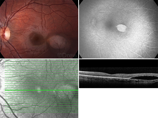
Torpedo Maculopathy
Written by Kevin C. Firl, MS, and Sandra R. Montezuma, MD, Department of Ophthalmology & Visual Neuroscience, University of Minnesota, Minneapolis; photos by Drew Miller, University of Minnesota Health, Minneapolis

A 3-year-old girl with a history of partial biotinidase deficiency diagnosed during infancy was referred to us for evaluation of a retinal lesion in her left eye. She was being followed by a pediatric metabolism clinic provider and was taking biotin for her biotinidase deficiency, and she was asymptomatic. Her visual acuity was 20/20 in both eyes, and she had an unremarkable anterior segment examination. However, the fundus exam of the left eye revealed a hypopigmented, oval-shaped lesion temporal to the macula of approximately 1 disc diameter. The lesion was longer in the horizontal axis than in the vertical axis, and it had a head pointing toward the macula. Optical coherence tomography (OCT) showed a large, subretinal cleft with attenuation of the retinal pigment epithelium (RPE) and outer retinal layers. Fluorescein angiography (FA) showed a window defect of the lesion with no leakage.
This presumably congenital lesion with a window defect on FA, attenuated RPE on OCT, and a head pointing toward the macula is consistent with torpedo maculopathy, a benign condition that requires no treatment. Other congenital retinal lesions, such as those seen in congenital hypertrophy of the RPE and familial adenomatous polyposis (Gardner syndrome), may look similar, but differences—including a thickened RPE on OCT—would be expected. Although biotinidase deficiencies are known to be associated with visual disturbances, their association with retinal pigment changes, including torpedo maculopathy, is unclear.
Read your colleagues’ discussion.
| BLINK SUBMISSIONS: Send us your ophthalmic image and its explanation in 150-250 words. E-mail to eyenet@aao.org, fax to 415-561-8575, or mail to EyeNet Magazine, 655 Beach Street, San Francisco, CA 94109. Please note that EyeNet reserves the right to edit Blink submissions. |