Blink
Multiple Myeloma Presenting With Roth Spots
By Robin A. Vora, MD, and Sophia Y. Wang, MD, and photographed by Pamela Neal, The Permanente Medical Group, Oakland, Calif.
Download PDF

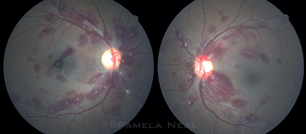
A 43-year-old man presented with flashes in his vision. His visual acuity was 20/70 in the right eye and 20/25 in the left. Dilated fundus exam revealed hemorrhages in multiple retinal layers and numerous Roth spots in both eyes. These spots may occur in many systemic conditions, including malignancies.
A stat complete blood count revealed 16,000/µL white blood cells, hemoglobin of 4.3 g/dL, and 94,000/µL platelets. The patient was admitted for transfusion. A bone marrow biopsy revealed hypercellular marrow composed mainly of plasma cells. Elevated serum lambda free light chains and an IgG spike on serum protein electrophoresis and immunofixation with quantitative protein level of 6.8 g/dL led to the diagnosis of multiple myeloma. Chemotherapy with cyclophosphamide, bortezomib, and dexamethasone was initiated.
| BLINK SUBMISSIONS: Send us your ophthalmic image and its explanation in 150-250 words. E-mail to eyenet@aao.org, fax to 415-561-8575, or mail to EyeNet Magazine, 655 Beach Street, San Francisco, CA 94109. Please note that EyeNet reserves the right to edit Blink submissions. |