Download PDF
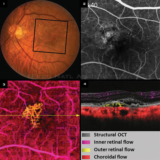
Fourier-domain optical coherence tomography (FD-OCT), the technology that already provides unprecedented views of the eye’s inner microstructures, is wowing ophthalmic researchers again—this time as a fast, noninvasive, 3-D alternative to fluorescein angiography (FA). Thanks to a special processing algorithm developed for existing high-speed OCT devices, OCT angiography can produce images of capillary-level blood flow in the retina and choroid in a few seconds, a team of Oregon researchers reported this spring.1
No dye required. “It’s a lot faster and more user-friendly than fluorescein angiography,” said David Huang, MD, PhD, study investigator and Peterson Professor of Ophthalmology at the Casey Eye Institute of Oregon Health & Science University, in Portland.
“Unlike fluorescein angiography, OCT angiography uses the motion of blood cells as contrast. So there is no dye injection, and you don’t have to take images at precise seconds over 10 minutes. There is no dye leakage and staining to obscure the boundary of pathologies. And, whereas with fluorescein you can only capture early transit in one eye, with OCT you can take your time and scan the other eye, too,” he said.
 |
|
PATIENT WITH AMD. (1) Black square indicates area of angiograms. (2) Late-stage fluorescein angiography showing an occult CNV. (3) Composite en face color-coded OCT angiogram with CNV flow highlighted in yellow. The CNV area was 0.96 mm2. Dashed line indicates the position of the OCT cross-section. (4) Cross-sectional color OCT angiogram; CNV is predominantly under the RPE. (Color key applies to both Figs. 3 and 4.)
|
New algorithm overcomes problems. The use of OCT for angiography has been studied since 2006, but early techniques required many repeat scans at the same location, a process too slow to be clinically practical, Dr. Huang said. “Also, people were not separating circulations in different layers, so a pathology like choroidal neovascularization could be obscured by retinal and choroidal vascular networks,” he said.
In their study, Dr. Huang and colleagues reported solving these problems with a split-spectrum amplitude-decorrelation angiography algorithm (SSADA). It can detect speckle variation in narrow spectral channels to optimize the detection of motion, he said. SSADA can produce a high-quality angiogram by repeating scans only once at each location. So the entire macula (6 × 6 mm) can be captured in 3 seconds with a new-generation commercial OCT system, Dr. Huang said.
Separate layers revealed. “Using en face visualization of separate layers in the retina and choroid, the algorithm can clearly visualize retinal and choroidal capillary networks and quantify capillary dropout, as well as measure retinal and choroidal neovascularization in normally avascular layers,” Dr. Huang said. “These clean images of pathology have gotten people really excited.”
The group has also had success identifying retinal pathology that is too early to see clinically, he added. “We have a diabetic case where there was a tiny dot of leakage that looked like a microaneurysm on FA, and it turned out on OCT angiography to be in the vitreous. So it is actually an early tuft of neovascularization.”
Limitations. According to the authors, the limitations of this technology include projection of the retinal flow signal onto deeper layers, such as the retinal pigment epithelium and choroid, and a relatively small field of view unless several scans are montaged.
Availability. Interest in OCT angiography mushroomed over the last year as angiography-enabled OCT systems entered commercial use internationally, Dr. Huang said. European and Asian sales of the 70-kHz Avanti system (Optovue), containing the Oregon group’s SSADA software, began in late 2014. Competitors also have begun adding other versions of angiography software to their OCT devices, he said.
—Linda Roach
___________________________
1 Jia Y et al. Proc Natl Acad Sci U S A. 2015;112(18):E2395-E2402.
___________________________
Relevant financial disclosures—Dr. Huang: Carl Zeiss Meditech: P; Optovue: O,P,S.
For full disclosures and disclosure key, see below.

More from this month's News in Review