Download PDF
It’s not every day that ophthalmologists save lives. But the eyes may be a proverbial canary in the coal mine for obstructive sleep apnea (OSA)—the most common type of sleep-disordered breathing, which increases the overall risk of death one and a half times in serious cases (patients who scored 30 or more on the apnea-hypopnea index).1
About 1 in 4 men and 1 in 10 women have sleep-disordered breathing. Yet most cases remain in the dark, undiagnosed.1 Despite its growing recognition by sleep specialists, OSA has not gained much traction in the field of ophthalmology, said Nancy M. Holekamp, MD, vitreoretinal surgeon and associate professor of clinical ophthalmology and visual sciences at Washington University, in St. Louis. “But ophthalmologists have a big role to play and can make a meaningful improvement in people’s lives.”
Deepak P. Grover, DO, a neuroophthalmologist practicing near Philadelphia, agreed. “The ophthalmologist may be able not only to detect ocular pathology associated with OSA at a stage early enough to maintain and restore vision but also to refer appropriately and help in the management and prevention of well-known systemic comorbidities such as coronary artery disease.”
OSA: Taking Your Breath Away
In patients with OSA, the musculature of the airway relaxes excessively during sleep, said Jamie M. Bigelow, MD, sleep specialist with the California Center for Sleep Disorders in the San Francisco Bay Area.
Under pressure. “The intake of breath creates a negative pressure that pulls in the walls of the airway, obstructing or narrowing it, and causing interruptions in breathing up to a hundred times an hour,” she said. As a result of these obstructing events, oxygen saturation may also be intermittently reduced.
All about oxygen. “Recent investigations suggest that the severity of oxygen desaturation may be a stronger correlate with cardiovascular problems than the actual number of breathing interruptions,” said Dr. Bigelow. “The drop in oxygen appears to unleash a whole host of changes, including release of catecholamines and inflammatory cytokines, which may play a role in injury and imperfect repair of blood vessels.” Patients with sleep apnea have been shown to have a higher incidence of hypertension, stroke, myocardial infarction, arrhythmias, diabetes, and dementia, said Dr. Bigelow, and in most cases, sleep apnea treatment has been associated with decreased risk or clinical improvement. As for the impact on the eye? Yo-yo-ing oxygen levels have untold effects over the long term, said Dr. Holekamp.
|
Is It Apnea?
|
 |
|
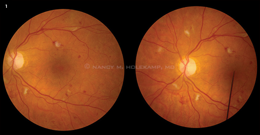
Fundus photos reveal multiple cotton-wool spots in a diabetic patient with suspected sleep apnea.
|
Ocular Diseases Linked to Sleep Apnea
Just as it’s important for ophthalmologists to be alert to hypertension or mild diabetic retinopathy, it’s also critical to recognize visual conditions that might be associated with sleep apnea, said Karl C. Golnik, MD, neuro-ophthalmologist at Cincinnati Eye Institute. Dr. Grover suggests having a high suspicion of sleep apnea if patients with predisposing factors (see “The OSA Profile”) present with any of the following five ocular conditions.
Floppy eyelid syndrome. This condition is Dr. Grover’s number-one reason for referring patients for a sleep study. One theory to explain floppy eyelid syndrome is a weak tarsal plate, common in obese patients; another involves the central nervous system. Normally, a person would be awakened by the sensation of pressure from pillows or bedding on an open eye, but “in patients with sleep apnea, a decrease in cortical arousability causes the eyelid to remain open when disturbed by mechanical stress during sleep,” he said. Over time, the lid becomes more lax and is easily everted with slight lateral traction.
Dr. Grover recommends referring patients with signs of lid laxity, especially men with other OSA risk factors, for a possible sleep study—even before full-blown signs of floppy eyelid syndrome appear. Topical treatment may help prevent papillary conjunctivitis and minimize symptoms such as dry eye, burning, and irritation.
NAION. Nonarteritic anterior ischemic optic neuropathy (NAION) is another strong reason for referral, said Dr. Grover, explaining that in several large studies, 70 to 80 percent of patients with NAION have been found to have OSA. What originally prompted investigation into the links between NAION and sleep apnea, said Dr. Grover, is the classic presentation of acute painless vision loss upon awakening in the morning in 75 percent of NAION patients. Although it is not possible to reverse vision loss from NAION, he said, treatment for sleep apnea may help prevent an attack of NAION in the other eye, which occurs in 15 to 18 percent of cases.
Papilledema. Linked to idiopathic intracranial hypertension (IIH), which occurs most frequently in young women, papilledema may be associated with increased venous blood flow, said Dr. Golnik. An increase in CO2 concentration may result from interrupted breathing, he said, and it may dilate blood vessels and increase pressure, leading to optic disc swelling.
Dr. Golnik advocates questioning all papilledema patients about symptoms of sleep apnea. He sends patients who report symptoms for a sleep study, as well as those who don’t fit the usual IIH demographic, such as men or anyone over age 50. Dr. Golnik recently referred an IIH patient for evaluation and treatment, which improved her vision and papilledema within a matter of weeks. The sleep doctor was incredulous, asking, “How did you know? She had some of the worst apnea I’ve ever seen. You saved her life.”
Glaucoma. A number of studies have examined the possible connections between OSA and glaucoma, but they have yielded varying results. For example, a large chart review of 156,336 patients with a diagnosis of sleep apnea initially showed an increased risk of open-angle glaucoma (OAG), but the difference disappeared with multivariable analysis that accounted for confounding factors.2 In contrast, other researchers have shown associations, including a 2012 study that found a link not only to primary OAG but also to ocular hypertension. Glaucoma patients with OSA had a higher intraocular pressure (IOP), worse visual field indices, and thinner retinal nerve fiber layer compared with the control group.3
Retinal conditions. Studies suggest a causal relationship between central serous chorioretinopathy (CSCR) and OSA, said Dr. Grover, because of the known increase in catecholamines with OSA. “Although CSCR can resolve within six months of [ophthalmic] treatment, sleep apnea treatment in patients with the condition has been shown to accelerate the recovery,” he said, citing a case of bilateral CSCR in which the patient’s vision returned to 20/20 and 20/25 and the serous detachment resolved within a week of starting apnea treatment.4
Causing severe dysfunction in the autoregulation of three major blood vessels—the posterior ciliary, central retinal, and ophthalmic arteries—OSA-related hypoxia may be a culprit in retinal vein occlusions, said Dr. Grover. Hypoxia is also the primary stimulus for neovascularization in diabetic retinopathy, he said.5 In addition, OSA’s potential role in diabetic retinopathy was spotlighted in a recent Oxford study, which found a high prevalence of sleep apnea in patients with diabetic clinically significant macular edema (CSME).6
“When your retina doesn’t get enough oxygen,” said Dr. Holekamp, “this adds insult to injury, exacerbating existing underlying problems like diabetic retinopathy or hypertensive retinopathy. The tip-off is six or more peripapillary cotton-wool spots [Fig. 1]. Clinicians traditionally call this hypertensive retinopathy, but it may be a manifestation of blood pressure spikes from obstructive sleep apnea. Nothing is 100 percent, but I’m batting a thousand with the diabetic patients with cotton-wool spots I’ve referred for sleep studies.”
The OSA Profile
The National Heart Lung and Blood Institute and the American Sleep Association list the following risk factors for, and signs and symptoms of, obstructive sleep apnea.
RISK FACTORS
Obesity
Large neck size
Enlarged tonsils in children
Small airway due to nasal congestion or bony structure
Family history of sleep apnea
Increasing age
Male gender
African-American, Hispanic, or Pacific Islander ethnicity
SIGNS AND SYMPTOMS
Loud snoring
Gasping or choking while asleep
Frequent nighttime urination
Morning headaches, dry mouth, or sore throat
Lack of energy or excessive daytime sleepiness
Hypertension
Memory, learning, or concentration problems
Depression, irritability, or mood swings
|
Spotting OSA-Related Eye Problems
Ophthalmologists can spot signs of visual conditions that may be linked with sleep apnea, said Dr. Golnik. “There’s no great secret here. A dilated retinal exam will catch many.” In some cases, he said, it may be an incidental finding or, in the case of NAION, patient-identified sudden vision loss.
Screening for sleepiness. Is the patient falling asleep while reading, watching television, at the wheel? Tired first thing in the morning? For any eye condition linked with sleep apnea, said Dr. Golnik, you can make a case for using a tool such as the Epworth Sleepiness Scale or Berlin Questionnaire—except when your suspicions are so high that it makes sense to go straight to a sleep study.
Dr. Holekamp finds these tools, which depend on patient responses, less reliable for her diabetic patients than doing a retinal exam. “Patients aren’t particularly good historians,” she said. “If they’ve had apnea for a long time, they may have developed compensatory mechanisms and not realize how tired they are all the time.”
Ordering a sleep study. Dr. Grover will first inquire about symptoms of excessive daytime somnolence and snoring or breathing difficulties, which may be reported by a spouse. “If my suspicion is very high, I’ll go ahead and order a sleep study and refer the patient to a sleep specialist.” In cases of mild to moderate suspicion, he will refer the patient to a primary care physician for an evaluation first and for consideration of unattended home-based portable polysomnography, which is less expensive than in-laboratory polysomnography.
Gray areas remain, however. For example, given the numbers involved, should all glaucoma patients be screened for sleepiness or referred for a sleep study? And what about patients with high blood pressure—is OSA a contributory factor? Maybe screening for OSA should be part of the history in these cases, said Dr. Holekamp, “but perhaps it should be a part of review of systems for everybody.”
 |
|
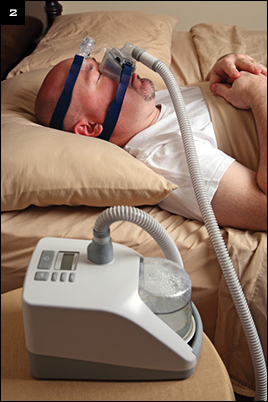
CPAP. Sleep apnea patient using continuous positive air pressure device.
|
Effects of OSA Treatment
Sleep apnea is highly treatable, and many of its symptoms—including ocular effects—are reversible, as long as the patient adheres to the regimen, said Dr. Grover. What’s more, added Dr. Bigelow, blood vessels remodel in response to treatment within three to four months, normalizing risks for strokes and heart attacks.
OSA treatment is based on the severity of the condition, patient preference, and affordability, said Dr. Bigelow. “For more severe cases, you need greater pressure to maintain patency of the airway. Continuous positive airway pressure (CPAP) is typically prescribed, but insurance may not cover it for milder cases.”
CPAP. Introduced in the 1980s, CPAP delivers pressure to the upper airway to prevent the collapse of the pharynx (Fig. 2). “CPAP does wonders,” said Dr. Grover. “My patients come back and say, ‘I feel more alive than I ever have.’” Dr. Holekamp sees cotton-wool spots disappear in her patients with diabetes. “With treatment, one patient was able to stop two blood pressure medications and one diabetes medication.”
But even though it can work well, the CPAP device can take some getting used to, said Dr. Golnik. Ocular side effects, such as irritation and tear evaporation, can also be an issue. Proper mask fit and optimal oxygen titration can resolve many of these problems, said Dr. Grover. He cautioned that some data indicate that treatment with CPAP may actually increase glaucoma patients’ nocturnal IOP, so these individuals need close monitoring.
Alternatives to CPAP. One alternative to CPAP is bilevel positive airway pressure (BiPAP), which allows independent adjustment of pressure during inhalation and exhalation.
Newer FDA-approved approaches include one-way valves placed in the nostrils to increase nasal expiratory positive airway pressure (Provent; Ventus Medical) and a device that applies vacuum through a mouthpiece to pull the soft palate forward (Winx; Apnicure).
Other options. Some patients may see improvement in their OSA with weight loss and avoidance of smoking, alcohol, and sedatives. Positional therapy or custom-made dental appliances that move the jaw forward are other options, said Dr. Bigelow.
Surgery—which may involve stiffening or shrinking tissue or removing the tonsils, uvula, and part of the soft palate—should be considered only as a secondary option, said Dr. Bigelow, because the procedures are quite invasive and the results are variable.
___________________________
Drs. Bigelow, Golnik, Grover, and Holekamp report no related financial interests.
___________________________
1 Punjami NM et al. PLoS Med. 2009;6(8):e1000132.
2 Stein JD et al. Am J Ophthalmol. 2011;152(6):989-998.e3.
3 Moghimi S et al. Sleep Med. 2012 Sep 1. [Epub ahead of print].
4 Jain AK et al. Graefes Arch Clin Exp Ophthalmol. 2010;248(7):1037-1039.
5 Ferrara N. Am J Physiol Cell Physiol. 2001;280(6):C1358-C1366.
6 Mason RH et al. Retina. 2012;32(9):17911798.