By Linda Roach, interviewing Kenneth L. Cohen, MD, Jeffrey L. Marx, MD, Safeer F. Siddicky, MS, and Scott E. Olitsky, MD
Download PDF
More than a decade after first being spotlighted at an Academy annual meeting, work-related musculoskeletal disorders (MSDs) remain a problem that must be solved one ophthalmologist at a time. According to Jeffrey L. Marx, MD, who began calling attention to the problem in 2001, there is still substantial interest in the topic. Indeed, a special session on ergonomics at AAO 2017 drew a standing-room-only crowd.
Dr. Marx, a vitreoretinal specialist in Massachusetts, noted that interest is particularly growing among younger ophthalmologists: “We had more YOs in the room than ever before—and I think that is both good and bad.
“It’s a reflection of their interest in trying to keep themselves healthy throughout their career. But, unfortunately, the bad news is that even the younger ophthalmologists are being affected by the significant burdens that we see in our clinical lives—seeing more and more patients and perhaps being at greater risk over time because of those increased burdens of everyday practice,” he said.
Dimensions of the Problem
Certain types of movements and tasks that are routine in ophthalmology can lead to cumulative MSDs of the back, shoulders, neck, and upper extremities, ergonomics experts say. Risk factors include:
- Repetitive tasks, especially under stressful circumstances.
- Tasks that require fine motor control and close visual focus. These increase muscular tension in the head, neck, and upper extremities.
- Prolonged maintenance of awkward body positions while working.
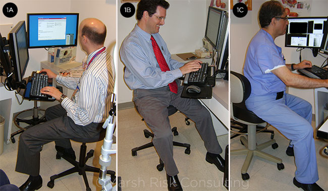
- Use of computer keyboards for extended time periods, especially if back and wrist support are lacking or the monitor is poorly placed (Figs. 1A-1C).
Dr. Marx and colleagues at the Lahey Clinic Medical Center in Burlington, Massachusetts, published 2 papers in 2005 about their groundbreaking research on the problem.1,2 Their survey of clinicians around the country found that half of the 697 respondents (51.8%) reported having neck, upper extremity, or lower back symptoms. Since then, several surveys in the United States and abroad have reported similar findings.
Dr. Marx said he views the steady increase in the number of attendees at his annual meeting presentations as a barometer of a continuing problem. “At these ergonomic symposia, usually we spend 45 minutes or an hour in an after-meeting, where our colleagues from around the country are asking questions or sharing their suggestions for ways to make practices ergonomically safer,” he said. “I think I learn something every time.”
 |
|
COMPUTER WOES. These photos depict common problems related to computer use in the clinic. (1A) Neck twisting, keyboard and seat too high, pressure on hips and lower back. (1B) Slouching, keyboard too high, legs don’t fit under console, pressure on hips and lower back. (1C) Keyboard and seat too high, no back support, pressure on hips and lower back.
|
Seeking Data on Risks and Solutions
Scientific studies to measure the strain that repeated motions and awkward postures place on the body have been conducted largely for manual occupations such as manufacturing and assembly lines, not ophthalmology. Nor are there objective metrics for determining whether purportedly “ergonomic” design features of new equipment actually reduce muscular tension and/or risks for users, said Scott E. Olitsky, MD, a pediatric ophthalmologist at the University of Missouri and Children’s Mercy Hospital in Kansas City, Missouri.
Analyzing the problem. “One of the things we really need to do is find ways to measure all of the angles we hold with our necks and backs throughout a procedure and quantify whether a new technique or tool is better or not,” Dr. Olitsky said. “What makes it ergonomic? Is there really data to determine that this desk or that chair or any other piece of equipment is ergonomically appropriate?”
Such questions are not just academic for Dr. Olitsky, who had to stop clinical and surgical practice 4 years ago after developing cervical radiculopathy.
Dr. Marx agreed that a more objective approach to ophthalmic ergonomics is needed. “We’ve never really advanced the science of ergonomics in ophthalmology,” he said. “We’ve qualitatively described the issue, and quantitatively described that there’s a problem, in terms of the percentages of ophthalmologists who, on surveys, say they have symptoms. But we haven’t really understood the science truly behind it.”
Insights from motion capture. The handful of nonsurvey studies that have been published were based on using electrogoniometry (which measures angles of joints) or inclinometers to track deviations of posture from neutral, and electromyography to measure muscle loading, in both clinical and surgical settings.3,4
Most recently, however, Dr. Olitsky and colleagues at the University of Missouri have begun studying ophthalmologists in action through motion-capture technology, similar to that used in Hollywood to bring lifelike movement to digital characters in movies.
The new system consists of a motion-capture suit, dotted with reflective markers, and 14 infrared video cameras that track the markers’ locations 3 dimensionally in space as the wearer moves, said Safeer F. Siddicky, MS, a doctoral student who serves as the mechanical engineer on the research team.
The researchers reported the results of their pilot study last November at AAO 2017.5 In the study, 10 pediatric ophthalmologists, outfitted in the motion-capture suit, were monitored to objectively determine how much their necks deviated from neutral during simulated retinoscopy and refraction, performed on an upright and then reclining mannequin.
Study findings and implications. The study found that during loose-lens retinoscopy, the percentage of procedural time with nonneutral neck flexion (mean ± standard error of the mean) was 81.39% ± 2.57% when the mannequin was upright. This decreased to 69.45% ± 3.91% (p = .038) with the mannequin reclined. The only other statistically significant difference in the mean percentage of nonneutral neck flexion was between loose prism and prism bar refraction: 66.54% ± 3.80% vs. 74.57% ± 1.38% (p = .028), respectively.
Although it was a small pilot study and limited to pediatric ophthalmologists, the findings objectively confirmed a long-standing belief among those concerned with ophthalmic ergonomics: Small alterations in work routines can make a big difference. “Simple postural alterations (such as reclining the patient during retinoscopy and refraction exams) may reduce the time spent by ophthalmologists in nonneutral postures, reducing the likelihood of musculoskeletal injuries,” the researchers wrote in their Academy poster.5
This cutting-edge type of motion analysis might eventually help the broader ophthalmology community better understand how to limit their MSD risks by modifying their work habits, Dr. Marx said.
“Most industries have used these types of studies to increase efficiency and decrease risks for their workers,” he said. “I think it could be a great advance for this field to understand what repetitive motions are absolutely necessary and what are probably unnecessary—and that we’re not even aware that we’re doing.”
Challenges for the Future
More time at the computer. With the growing use of electronic health records, it is becoming increasingly important for ophthalmologists to pay attention to the ergonomics of how they document patient visits. More time at a computer keyboard or manipulating a mouse could lead to MSDs of the hands, arms, neck, and back, if the exam room setup prevents the clinician from arranging the chair, keyboard, mouse, and monitor properly, Dr. Marx said. Experts say the monitor should be at or slightly below eye level; forearms should be angled only slightly downward; and a chair with armrests and good back support should be used.
Heads-up displays in the OR. The operating microscope has been linked to neck problems among surgeons, and heads-up displays are being viewed as a possible solution. However, this presumes that the monitor’s position can be adjusted to the surgeon’s stature so that the neck is not flexed or extended when viewing it, Dr. Olitsky said. “A good tool isn’t a good tool unless it’s installed correctly,” he noted. In addition, an assisting surgeon should avoid twisting the back and neck to view a monitor being used by the primary surgeon, he said.
Cramped operating rooms. A proliferation of devices in the ophthalmic operating and procedure rooms is making them more crowded than ever, which can create difficulties for surgeons attempting to heed ergonomic advice, said Kenneth L. Cohen, MD, who is the Sterling A. Barrett Distinguished Professor of Ophthalmology at the University of North Carolina.
“The operating room has become a more complex arena, and thus the physical nature of surgery requires attention to ergonomics,” Dr. Cohen said. “For example, there are more standalone instruments. There are lasers for retinal surgery, there are femtosecond lasers for cataract surgery, there are IOL positioning devices, [and] there are video monitors. The placement of these devices affects the surgeon at the microscope—hand position, foot pedal position, and, of course, patient position.”
Despite these challenges, surgeons should always adjust both the operating equipment and the patient bed in ways that keep their necks and backs aligned neutrally, Dr. Olitsky advised. Doing so is an investment not just in their health today but also in their long-term professional futures, he said. “We all sometimes think we can’t take the time to do this or do that. But the reality is that taking those few minutes now may greatly extend your career.”
___________________________
1 Dhimitri KC et al. Am J Ophthalmol. 2005;1391:179-181.
2 Marx JL et al. Techniques in Ophthalmology. 2005;31:54-61.
3 Fethke NB et al. Int J Ind Ergon. 2015;49:53-59.
4 Shaw C et al. Can J Ophthalmol. 2017;52(3):302-307.
5 Siddicky SF et al. Evaluating ergonomics in ophthalmology using kinematic motion analysis: a pilot study [PO386.] Poster presented at: AAO 2017; Nov. 17, 2017; New Orleans. (ePoster available at: aao.scientificposters.com.)
___________________________
Dr. Cohen is the Sterling A. Barrett Distinguished Professor of Ophthalmology at the University of North Carolina, in Chapel Hill. Dr. Marx is a vitreoretinal specialist at Lahey Medical Center in Burlington, Mass. Dr. Olitsky is Section Chief, Ophthalmology, at Children’s Mercy Hospital, and professor of pediatric ophthalmology at the University of Missouri–Kansas City (UMKC) School of Medicine. Mr. Siddicky is a PhD candidate in mechanical engineering and bioinformatics at UMKC. Relevant financial disclosures: None.
Further Reading:
Physician Wellness: How to Avoid the High Cost of Physician Burnout (EyeNet, November 2017)
Ophthalmic Ergonomics: Seven Risk Factors and Seven Solutions (EyeNet, September 2009)
Wellness Resources
Visit aao.org/wellness for a panoply of tools and information to help you reduce stress, avoid burnout, and promote well-being in your professional and personal life.
|