By Sheri L. DeMartelaere, MD, Sean M. Blaydon, MD, and John W. Shore, MD, FACS
Edited By Thomas A. Oetting, MD
This article is from September 2005 and may contain outdated material.
When I first met with Ann Smith,* she was a healthy 57-year-old woman with hypertension and mildly elevated cholesterol who was complaining of severely itchy eyelids. My initial reaction when seeing the history on the chart was that I would be dealing with another routine patient with blepharitis and dry eyes. That is, until I actually started my examination and suddenly realized that she had something I had never encountered before.
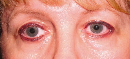
Ms. Smith’s upper and lower eyelids on both sides were thickened and inflamed. Her eyelid margins had indiscriminate scattered lash loss. There were elevated, firm, linear, nodule-like masses located diffusely along each eyelid margin. These masses were nontender, and I detected no regional adenopathy. Her vision was unaffected, and the remainder of her eye examination was unremarkable. As I examined her, Ms. Smith told me the itching was so severe she felt like “ripping [her] eyelids off.” Thankfully, she used cold compresses instead, which gave her some relief.
After mentally scolding myself for going right to the exam, a habit easy to fall into, I realized I needed to hear the “rest of the story.” Ms. Smith’s path to our office began several months ago when she first went to see her primary care doctor complaining of a two-week episode of itchy, swollen eyelids. These semiacute changes coincided with her starting atorvastatin (Lipitor). Her physician felt that her itchy eyelids might be an allergic reaction and so discontinued the atorvastatin. Because of the severity of her symptoms, her physician also prescribed a short course of oral steroids.
The symptoms initially improved, but two weeks after she completed the steroids Ms. Smith’s problems were back, and seemingly worse. She was in so much discomfort that she went to her local urgent care clinic. There, a different physician felt she was having an allergic reaction to her hydrochlorothiazide, and so it was also discontinued. She was started on irbesartan (Avapro) for her hypertension, given a steroid injection and placed on over-the-counter allergy eye drops.
One week later, after no improvement, she was given another course of oral steroids, switched to different eye drops, and referred to a dermatologist.
 |
|
What’s Your Diagnosis? Itchy, swollen lids with lash loss, inflammation and nodule-like masses.
|
Additional History Is Revealed
At the dermatologist’s office, Ms. Smith recalled that more than a year ago she had had permanent eyeliner tattooed on both upper and lower eyelids. She hadn’t mentioned this to her previous doctors because she was certain it couldn’t be the source of her problem. Before getting the tattoo, she had undergone a pigment patch test that read negative at one week. During her consult, the dermatologist performed a repeat patch test and took a biopsy of her left upper eyelid.
The new patch test revealed a 2+ reaction to the same pigment used to tattoo her eyelids. The eyelid biopsy revealed hyperkeratosis with chronic inflammation as well as granulomatous inflammation surrounding black pigment material.
Ms. Smith was diagnosed with a delayed hypersensitivity reaction to the eyeliner pigment and was referred to our office for definitive treatment. As she told me this, I knew I needed to hit the books.
A Treatment Dilemma
I learned that treatment for an allergic reaction to blepharopigmentation can be very challenging. Multiple treatment modalities have been tried, including topical steroid creams, local steroid injections, local resection, intramuscular steroid injections, systemic oral steroids and antihistamines.¹
Although laser is commonly used to remove erroneously placed pigment, laser treatment is not recommended in the allergy setting. The laser shatters the pigment and actually enhances antigen presentation to the immune system. And it doesn’t “erase” the tattoo; it simply creates finer particles for macrophages to engulf. Making the particles smaller doesn’t reduce their allergic propensity and instead runs the risk of causing systemic distribution of the pigment and concomitant systemic allergic issues.²
After consulting several other dermatologists and ophthalmologists, Ms. Smith was placed on fluorometholone ophthalmic ointment to her eyelid margins twice daily. After several months, her symptoms significantly subsided.
The same day that Ms. Smith came to see us, I saw an FDA Alert warning consumers about adverse events associated with certain shades of the Premier Pigment brand of tattoo ink. Side effects included inflammation, blistering, scarring and the formation of granulomas in the periorbital area.
Unfortunately, this is exactly the brand of pigment that Ms. Smith received. (The Premier pigments associated with adverse reactions are listed at this Web address: www.cfsan.fda.gov/~dms/cos-tat2.html.)
It is important to recognize that skin tests before blepharopigmentation will not predict a delayed hypersensitivity reaction.
We used to offer blepharopigmentation in our office. However, after seeing and treating Ms. Smith, we decided to discontinue that service.
Diagnostic Keys of “Blepharopigmentitis”
| Signs and Symptoms |
Differential Diagnosis |
How to Make the Diagnosis |
Intense
eyelid itching |
Staphylococcal marginal blepharitis
Contact dermatitis
Atopic dermatitis
Hyperpigmentation due to drugs |
Previous blepharopigmentation
Dermal patch test with controls
|
| Eyelid edema |
Blepharochalasis
Thyroid-associated
ophthalmopathy
Idiopathic orbital
inflammation
Angioneurotic edema
due to drugs |
Signs and symptoms localized to areas of pigment |
Erythema-
tous lid margins |
Preseptal cellulitis
Staphylococcal marginal blepharitis
Seborrheic blepharitis/rosacea
Hordeolum |
Previous blepharopigmentation |
Lid margin crusting/
blisters |
Herpetic blepharitis
Staphylococcal marginal blepharitis
Angular blepharitis
Exfoliative dermatitis due to drugs |
Previous blepharopigmentation
Dermal patch test with controls |
| Lash loss |
Neoplasia of any type, especially sebaceous cell carcinoma
Seborrheic blepharitis
Herpes zoster
Idiopathic/self-epilation
Trauma |
Biopsy reveals granulomatous inflammation withoutevidence of infectious organisms or carcinoma
No regional adenopathy |
Firm
nodular masses |
Neoplasia of any type
Multiple chalazia
Molluscum contagiosum
Sebaceous adenoma
Sarcoidosis |
Biopsy reveals granulomatous inflammation without evidence of infectious organisms or carcinoma
No regional adenopathy |
|
* Patient name is fictitious.
___________________________________
1 Vagefi, M. R. et al. Adverse reactions to permanent eyeliner tattoo. (Poster presented at ASOPRS Scientific Symposium, 2004.)
2 Schwarze, H. P. et al. J Am Acad Dermatol 2000;42:888–891.
___________________________________
Dr. DeMartelaere is an ophthalmic plastic and reconstructive surgery fellow who is working with Dr. Shore at Texas Oculoplastic Consultants, Austin, Texas. Dr. Blaydon is also on staff at Texas Oculoplastic Consultants.