Download PDF
Using a new imaging technique, researchers for the first time have visualized retinal cell death in human subjects.1 Their proof-of-concept study involved glaucomatous eyes, but the technique, known as DARC (detection of apoptosing retinal cells), has implications for observing cell death in other neurodegenerative diseases and may eventually play a role in predicting early neurodegenerative activity.
“It is a novel technique for looking at ‘sick’ single nerve cells, which indicate disease activity,” said M. Francesca Cordeiro, PhD, MRCP, FRCOphth, at the Imperial College London.
 |
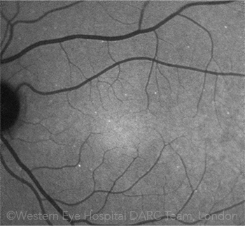
CELL DEATH. This image of a patient’s retina shows hyperfluorescent signals. Each white spot is a single sick retinal nerve cell, labeled in vivo with the DARC technique.
|
Counting sick cells. This phase 1 clinical trial involved 8 patients with glaucomatous neurodegeneration and evidence of progressive disease, plus 8 healthy controls. All received intravenous ANX776 (annexin 5, a cellular protein that binds to an apoptosis marker, compounded with fluorescent dye DY-776).
Retinal angiograms and optical coherence tomography images were acquired before, during, and after injection. Apoptotic cells appeared as unique hyperfluorescent spots on the retina. The number of spots, or the DARC count, was significantly higher (2.37-fold) in glaucoma patients, compared with healthy controls.
In other findings, the DARC count significantly correlated with decreased central corneal thickness and increased cup-disc ratios in glaucoma patients. In healthy controls, the DARC count was correlated with increased age. In addition, no adverse effects were noted in either the glaucoma patients or the healthy controls.
Surprise finding. The DARC count was greater in patients who later showed increasing rates of disease progression in any parameter—rim area, retinal nerve fiber layer, or visual field. “Increased DARC activity was predictive of increased structural and/or functional rates of progression,” Dr. Cordeiro said of the unexpected post hoc finding.
Up next. The predictive nature of the technique has potential clinical implications. DARC might help identify glaucoma suspects more quickly, said Dr. Cordeiro. It also might help researchers assess response to treatment.
Phase 2 studies will assess DARC in age-related macular degeneration, optic neuritis, and Alzheimer disease. Dr. Cordeiro is also developing a noninvasive DARC compound. “We believe a less-invasive delivery system would allow for more widespread screening and diagnostic testing,” she said.
—Miriam Karmel
___________________________
1 Cordeiro MF et al. Brain. 2017. Published online Apr. 26, 2017.
___________________________
Relevant financial disclosures—Dr. Cordeiro is a named inventor on DARC technology patents owned by University College London. The project has been funded through the Wellcome Trust charity through an academic pathway.
For full disclosures and disclosure key, see below.

More from this month’s News in Review