Download PDF
Retinal microvascular abnormalities and impaired blood flow appear to be emerging as signature ocular manifestations of COVID-19 infections, researchers have found.1,2 The abnormalities, which have been observed even in otherwise healthy eyes, support the hypothesis of widespread microvascular damage that could be clinically silent in those who have had COVID.2
 |
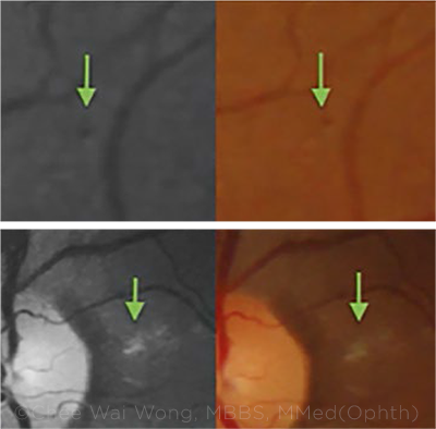
AFTER COVID. Retinal abnormalities, as seen on color fundus and red-free images, included (top) retinal microhemorrhages and (bottom) peripapillary cotton-wool spots (green arrows).
|
Study in Singapore. In Singapore, a prospective study of 108 COVID-positive subjects found that 1 in 9 had retinal microvascular signs.1 The abnormal findings included eight eyes (3.7%) with microhemorrhages, six (2.8%) with retinal vascular tortuosity, and two (0.93%) with cotton-wool spots. OCT scans revealed 11 eyes (5.1%) with hyper-reflective plaques in the ganglion cell-inner plexiform layer—and two of those eyes also had retinal signs visible on color fundus photographs.
Underlying cardiovascular impact? “These signs were observed even in asymptomatic patients with normal vital signs and no past history of cardiovascular disease. These retinal microvascular signs could represent underlying cardiovascular and thrombotic alterations associated with COVID-19 infection,” said Chee Wai Wong, MBBS, MMed(Ophth), at the Singapore National Eye Centre and the Duke-NUS Medical School.
Moreover, the scientists found that COVID patients with retinal signs were significantly more likely to have elevated blood pressure than were those without such signs (p = .03), Dr. Wong said. “Serial monitoring of these patients showed normalization of blood pressure as they recovered from COVID-19. Hence, there may be a role in triaging patients for eye screening based on serial monitoring of blood pressure,” he said.
Study in Italy. Italian researchers performed OCT and OCT angiography (OCTA) on the retinas of 40 patients who had recovered from SARS-CoV-2 pneumonia and compared them to 40 healthy control subjects.2
All of the post-COVID patients had normal ocular and fundus examinations, and none reported eye symptoms. Nonetheless, the OCTA images showed significant, diffuse perfusion loss in several areas of the post-COVID patients’ retinas, compared to controls, the researchers reported.
Alterations in retinal blood flow. The density of deep capillary vessels was reduced in all macular regions, as was vessel density in the superficial capillary plexus (p = .038) and the radial peripapillary capillaries (p < .001). Structural OCT showed lower average thickness of the retinal nerve fiber layer (p = .012), and this correlated significantly with the angiographic data.
Alterations in retinal blood flow could reflect a variety of pathogenic mechanisms that have been linked to SARS-CoV-2 infection, including thromboinflammatory microangiopathy and angiotensin-converting enzyme 2 disruption, the authors said.
If the group’s findings are confirmed by larger studies, OCTA images might help physicians to better anticipate or prevent systemic deterioration in their COVID patients, the researchers wrote. “OCTA allows [physicians] to detect the signs of retinal thrombotic microangiopathy that could reflect the systemic vascular impairment occurring in multiorgan dysfunction. OCTA could represent a valid biomarker of systemic vascular dysfunction after SARS-CoV-2 infection.”
—Linda Roach
___________________________
1 Sim R et al. Br J Ophthalmol. 2021;227:182-190.
2 Cennamo G et al. Am J Ophthalmol. Published online March 26, 2021.
___________________________
Relevant financial disclosures—Dr. Wong: None.
For full disclosures and the disclosure key, see below.
Full Financial Disclosures
Dr. Crama None.
Dr. Gillmann None.
Dr. Mansouri Allergan: S; ImplanData: C; Santen: C; Sensimed: C.
Dr. Ramanathan None.
Dr. Wong None.
Disclosure Category
|
Code
|
Description
|
| Consultant/Advisor |
C |
Consultant fee, paid advisory boards, or fees for attending a meeting. |
| Employee |
E |
Employed by a commercial company. |
| Speakers bureau |
L |
Lecture fees or honoraria, travel fees or reimbursements when speaking at the invitation of a commercial company. |
| Equity owner |
O |
Equity ownership/stock options in publicly or privately traded firms, excluding mutual funds. |
| Patents/Royalty |
P |
Patents and/or royalties for intellectual property. |
| Grant support |
S |
Grant support or other financial support to the investigator from all sources, including research support from government agencies (e.g., NIH), foundations, device manufacturers, and/or pharmaceutical companies. |
|
More from this month’s News in Review