Blink
Retinal Astrocytoma
By Cristina Moreira dos Santos, MD, Inês Coutinho, MD, and Susana Teixeira, MD, and photographed by Susana Teixeira, MD, Hospital Prof. Fernando Da Fonseca E.P.E. Lisbon, Portugal
Download PDF

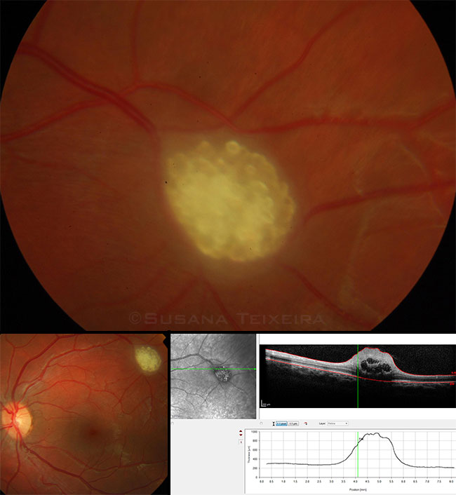
In 2003, a 3-year-old girl presented with a 0.5 disc diameter subretinal translucent lesion without vitreous seeding. Ultrasound showed no calcification. Because the appearance was not typical for retinoblastoma, serial funduscopic exams with photography were performed to track growth.
In the following years, spherical cysts resembling tapioca started to form. A presumptive diagnosis of retinal astrocytoma, a low-grade neoplasm of retinal origin, was established. Because retinal astrocytoma is often associated with tuberous sclerosis, a workup for tuberous sclerosis was performed, including tympanometry, renal and abdominal ultrasound, and brain CT scan and MRI. All results were negative.
The photos below were taken 12 years after first observation. The lesion is 1 disc diameter in size and comprises multiple cysts. SD-OCT shows a heterogeneous reflectivity of the lesion; thickening of the ganglion and inner plexiform layers; and, at the posterior, multiple hyporeflective cystic cavities. This peripheral lesion did not affect the patient’s vision.
| BLINK SUBMISSIONS: Send us your ophthalmic image and its explanation in 150-250 words. E-mail to eyenet@aao.org, fax to 415-561-8575, or mail to EyeNet Magazine, 655 Beach Street, San Francisco, CA 94109. Please note that EyeNet reserves the right to edit Blink submissions. |