Download PDF
A specialized imaging system developed at the NEI can directly visualize deterioration of the retinal pigment epithelium (RPE) over time—an achievement that is enabling the researchers to begin investigating the system’s utility for directly tracking the progression of blinding retinal diseases.
“We’ve only recently started to apply this technique to investigate diseases,” said study leader Johnny Tam, PhD, at the NEI. “Currently we’re interested in seeing how RPE cells are affected in diseases such as age-related macular degeneration as well as various inherited retinal degenerations.”
 |
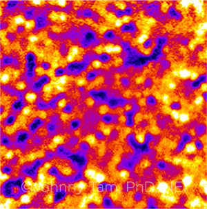
IN SITU. NEI scientists have visualized and tracked the mosaicism of RPE cells (shown here).
|
How it works. The imaging system combines adaptive optics (AO) with indocyanine green (ICG) angiography and scanning laser ophthalmoscopy to produce detailed structural images of the photoreceptor-RPE-choriocapillaris complex in living human eyes.1 (See “News in Review,” February.)
Focusing on the RPE. In their most recent study, the researchers concentrated their attention on the RPE. They injected ICG dye in healthy subjects with visual acuity of 20/20 or better and in patients with two types of progressive hereditary degenerative retinal diseases—late-onset retinal degeneration (L-ORD) and Bietti crystalline dystrophy (BCD).
The images from ICG injections taken months apart showed that the system could track changes in the RPE, the researchers found.2 The RPE cell mosaic was stable in the healthy eyes, was slightly less stable in L-ORD, and changed drastically in BCD.
“This study is a step toward functional imaging of RPE cells, in which we start to explore the dynamics of dye uptake and clearance across a large range of time—seconds to a year,” Dr. Tam said. “We believe that imaging the RPE, in combination with other clinical assessments, will allow us to identify patients who are at risk for losing their vision.”
Long-term goal. The research group’s long-term goal is to bring the lessons from its adaptive optics system into widespread clinical use, Dr. Tam said. As part of this, the researchers noted that they observed a characteristic AO-ICG fluorescence pattern in every healthy eye that they imaged, and they are using this information to create an in vivo database of human foveal RPE cell-to-cell spacing.2
“Translating this technique to a standardized clinical test is a tremendous endeavor, but we have achieved a critical first step by deploying our custom-built instrument in a clinical setting at the NEI’s Eye Clinic,” Dr. Tam said.
“In the past decade we’ve witnessed rapid advances in technology, and it would not be inconceivable to think that we can simplify this technique over the coming decade and make it robust enough to be used in a conventional clinical setting,” he said.
—Linda Roach
___________________________
1 Jung H et al. Commun Biol. 2018;1:189.
2 Jung H et al. JCI Insight. 2019;4(6):e124904.
___________________________
Relevant financial disclosures—Dr. Tam: None.
For full disclosures and the disclosure key, see below.
Full Financial Disclosures
Dr. Esmaeli None.
Dr. Ferdi Northcote Trust: S.
Dr. Rong None.
Dr. Tam None.
Disclosure Category
|
Code
|
Description
|
| Consultant/Advisor |
C |
Consultant fee, paid advisory boards, or fees for attending a meeting. |
| Employee |
E |
Employed by a commercial company. |
| Speakers bureau |
L |
Lecture fees or honoraria, travel fees or reimbursements when speaking at the invitation of a commercial company. |
| Equity owner |
O |
Equity ownership/stock options in publicly or privately traded firms, excluding mutual funds. |
| Patents/Royalty |
P |
Patents and/or royalties for intellectual property. |
| Grant support |
S |
Grant support or other financial support to the investigator from all sources, including research support from government agencies (e.g., NIH), foundations, device manufacturers, and/or pharmaceutical companies. |
|
More from this month’s News in Review