Download PDF
A recent analysis from the advanced Imaging for Glaucoma (AIG) study strengthens the case for using optical coherence tomography (OCT) in everyday clinical practice.1 However, the findings do not suggest that OCT will replace visual fields (VFs) at this time.
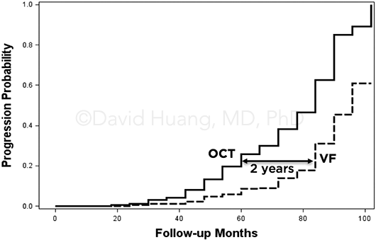
Early and late. The comparison of OCT and VF showed the usefulness of OCT structural analysis for detecting progression in both early and late stages of glaucoma, said David Huang, MD, PhD, at Casey Eye Institute in Portland, Oregon.
In early stages of glaucoma, peripapillary retinal nerve fiber layer (NFL) thickness and macular ganglion cell complex (GCC) thickness, as measured by OCT, were together more sensitive than were VF parameters in detecting progression. And in a separate finding, one Dr. Huang called “important and novel,” OCT detected progression in advanced glaucoma on a par with VF. The ability of OCT to monitor advanced glaucoma was mostly due to GCC, as the AIG study confirmed findings from other studies that showed NFL to be less sensitive in advanced disease.
 |
TREND ANALYSIS. Kaplan-Meier plots of glaucoma progression.
|
Multicenter analysis. The study, conducted at 5 universities, included 417 glaucoma suspect and preperimetric glaucoma eyes and 377 perimetric glaucoma eyes. Fourier-domain OCT was used to map the thickness of the NFL and GCC. OCT-based progression detection was defined as a significant negative trend for either average NFL or GCC thickness. VF progression was detected if either the visual field index (VFI) trend analysis or the Guided Progression Analysis (GPA) event analysis reached significance.
Findings. In the glaucoma suspect/preperimetric group, OCT detected progression in 38.9% of eyes, versus 18.7% using VF parameters. In early perimetric glaucoma, OCT had a significantly higher detection rate compared to VF: 49.7% versus 32%, respectively.
However, “In a significant percentage of eyes, progression was detected only on VF,” Dr. Huang said. “This percentage is large in later stages and small in the earlier stages of glaucoma.”
Clinical implications. Although OCT proved useful in monitoring progression, Dr. Huang said, “VF is needed in initial patient evaluation to establish the stage of glaucoma.” He added that in advanced disease, when patients need to be closely monitored, both technologies are needed.
Looking ahead. “The evidence is strong that OCT should be an important part of progression monitoring in preperimetric glaucoma and mild perimetric glaucoma,” Dr. Huang said. “Since OCT scanning is relatively quick, it could easily be performed on every visit. In early glaucoma, OCT could help catch progression sooner and measure the rate of progression more precisely.”
—Miriam Karmel
___________________________
1 Dastiridou A et al. Comparison of glaucoma progression detection by optical coherence tomography and visual field. Presented at: ARVO 2017 Annual Meeting; May 8, 2017; Baltimore.
___________________________
Relevant financial disclosures—Dr. Huang: Optovue: O,P,S.
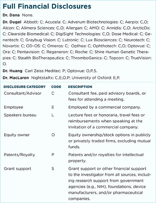
For full disclosures and disclosure key, see below.

More from this month’s News in Review