Blink
Susac Syndrome
By Carlos Perpétua, MD, Rita Couceiro, MD, Filipe Braz, MD, and Joaquim Prates Canelas, MD, Hospital De Santa Maria, Lisbon, Portugal
Photo by Carlos Perpétua, MD
Download PDF

A 26-year-old man presented with progressive and painless blurring of vision in the left eye. He had a 2-month history of hypoacusis, disorientation, memory loss, and behavioral changes. On examination, his best-corrected visual acuity was 20/20 in the right eye and 20/25 in the left.
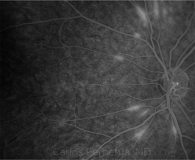
Funduscopy of the left eye revealed arteriolar narrowing and rarefaction. Fluorescein angiography showed delayed perfusion in both eyes, leakage of dye in several branch retinal arteries, and no perfusion distally, suggesting branch retinal artery occlusions (BRAO).
Audiometry revealed complete deafness of the right ear and neurosensory hypoacusis of the left ear. On brain magnetic resonance imaging, there were multiple focal white matter lesions above the tentorium and in the corpus callosum, with no contrast uptake.
The combined ophthalmic and systemic findings raised the suspicion of Susac syndrome. This is a rare disease of unknown origin, most likely an autoimmune endotheliopathy, causing an arteriolar microangiopathy of the brain, cochlea, and retina. Susac syndrome tends to affect young women, but it can also occur in men. It manifests as a clinical triad of subacute encephalopathy, hypoacusis, and visual loss caused by multiple BRAO.
| BLINK SUBMISSIONS: Send us your ophthalmic image and its explanation in 150-250 words. E-mail to eyenet@aao.org, fax to 415-561-8575, or mail to EyeNet Magazine, 655 Beach Street, San Francisco, CA 94109. Please note that EyeNet reserves the right to edit Blink submissions. |