Blink
Syphilitic Panuveitis and Retinitis in HIV
By Michelle C. Liang, MD, and Caroline Baumal, MD, and photographed by Jennifer Moran, New England Eye Center at Tufts Medical Center, Boston
Download PDF

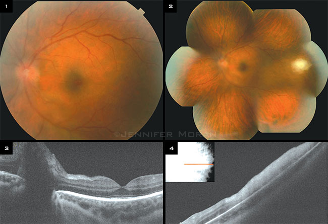
A 25-year-old man presented with a painful red right eye and decreased vision in both eyes for 1 week. Vision was 20/300 in his right eye and 20/50 in his left eye. Ophthalmoscopy revealed bilateral inflammatory cells in the vitreous, optic nerve swelling, and peripheral patches of retinitis, more severe in the right eye (Figs. 1, 2). Spectral-domain optical coherence tomography confirmed disc swelling and full-thickness retinitis in the temporal periphery of the left eye (Figs. 3, 4). Vitreous inflammation prevented imaging of the right eye.
Laboratory testing revealed a positive serum rapid plasma reagin, positive serum Venereal Disease Research Laboratory (VDRL), negative cerebrospinal fluid VDRL, and negative brain magnetic resonance imaging, consistent with a diagnosis of secondary syphilis.
Because of the ocular involvement, the patient was treated for neurosyphilis with IV penicillin for 3 weeks and intramuscular penicillin for 2 weeks. Systemic workup also revealed a new diagnosis of HIV, after which he was started on highly active antiretroviral therapy. Four months after presentation, the vitritis and retinitis were completely resolved. Vision was 20/50 in the right eye and 20/20 in the left eye.
| BLINK SUBMISSIONS: Send us your ophthalmic image and its explanation in 150-250 words. E-mail to eyenet@aao.org, fax to 415-561-8575, or mail to EyeNet Magazine, 655 Beach Street, San Francisco, CA 94109. Please note that EyeNet reserves the right to edit Blink submissions. |