Download PDF
Magical Clogs
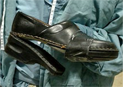
 Dr. Williams’ editorial on surgical rituals and magical thinking (Opinion, December 2016) got me thinking about my own equally irrational traditions. Since 2000, I’ve performed all of my hospital OR cases in the same pair of black clogs. Over the years, the leather uppers have separated from the rubber soles and were held together with (matching) duct tape. My fealty to them was less lucky rabbit’s foot and more security blanket. After all, they had been faithful companions to every single operative patient encounter, both routine and harrowing, throughout my surgical career. In December, my scrub remarked, “Dr. Fountain, we know what to get you for Christmas.” I told her not to bother, but I would appreciate some black duct tape in my stocking.
Just last week, however, I suffered a catastrophic wardrobe malfunction: The duct tape gave way, and I nearly tripped down the stairs. The damage was inoperable. I concluded that replacing my footwear, albeit with great reluctance, would be easier and less costly than replacing my hips. A brief solemn ceremony ensued where, interrupted by the muffled giggles of my 2 scrub nurses, I buried the mutilated leather and rubber in the trash bin of the operating room.
I’m breaking in a new pair of Danish clogs now. They’re shiny and still squeak a little with each footfall. I hope they’ll accompany me, like a loyal friend, through the end of my career. With any luck—and plenty of duct tape—they will.
Tamara R. Fountain, MD
Deerfield, Ill.
Blepharochalasis: A Cryptic Condition
“Blepharochalasis Syndrome” (Ophthalmic Pearls, September 2016) piqued my interest. Body skin has an average 1.75 m2 surface area, of which less than 20 cm2 is eyelid skin (0.1%). Nevertheless, this portion bears more than 5% of skinmalignancies; and due to its enormous cosmetic and functional values, the eyelid is a major medical and surgical field.
Edema occurs through hydrostatic pressure imbalance,as well as through a variety of mechanisms: vasogenic, inflammatory (including allergy), and autoimmune. The latter 3 have been suggested as the pathogenesis of the acute phase of blepharochalasis. There is pathologic and molecular evidence in support of inflammation, but the lack of response to steroids, antihistamines, and other anti-inflammatory agents substantially weakens the inflammation theory. I believe blepharochalasis to have an angioneurotic etiology and to be very much similar to angioedema, an underlying aberrant autonomic innervation and/or local dysautonomia.
Diagnosis of blepharochalasis is challenging, as it is a condition with no specific lab or imaging findings. The acute phases are transient and short-lived, so a history of episodes must be elicited by the physician and/or taken seriously when described spontaneously by the patient. Past blepharochalasis events should be differentiated from bouts of allergic reactions. And its episodic nature can be highly variable among individuals and throughout the course of the condition. However, in the chronic established case, the sequelae of hanging, atrophic, and telangiectatic skin are characteristic.
We take care of a tiny but dear portion of the body’s skin, and need to keep blepharochalasis in mind to avoid missing it. We should study and report more frequently on it to develop further evidence for its treatment.
S-Farzad Mohammadi, MD, MPH, FICO
Tehran, Iran
| WRITE TO US Send your letters of 150 words or fewer to us at EyeNet Magazine, American Academy of Ophthalmology, 655 Beach Street, San Francisco, CA 94109; e-mail eyenet@aao.org; or fax 415-561-8575. (EyeNet reserves the right to edit letters.) |