Blink
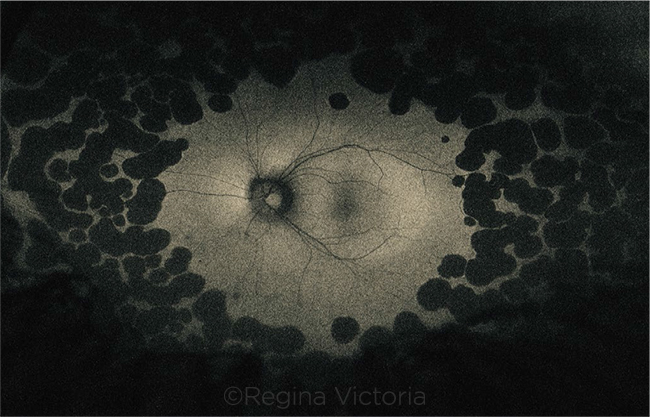
Widefield Fundus Autofluorescence in Gyrate Atrophy
By Miguel Paciuc-Beja, MD, and Hugo Quiroz-Mercado, MD
Photo by Regina Victoria, Denver Health Medical Center, University of Colorado School of Medicine, Aurora
Download PDF

A 16-year-old myopic girl presented with a complaint of diminished vision, especially at night. Current spectacle visual acuity (VA) was 20/60 in the right eye (OD) and 20/50 in the left eye (OS). Lensometry showed –8.25 OD and –8.75 OS. Refraction improved VA to 20/25 in both eyes with correction of –9.00 OD and –9.50 OS. Fundus exam showed well-demarcated, scalloped areas of outer retinal and inner choroidal atrophy in the retinal periphery that were hypoautofluorescent. Ornithine plasma levels were more than 10 times above the normal range (hyperornithinemia). A diagnosis of gyrate atrophy due to a deficiency of ornithine aminotransferase was made, and she was placed on a low arginine diet.
| BLINK SUBMISSIONS: Send us your ophthalmic image and its explanation in 150-250 words. E-mail to eyenet@aao.org, fax to 415-561-8575, or mail to EyeNet Magazine, 655 Beach Street, San Francisco, CA 94109. Please note that EyeNet reserves the right to edit Blink submissions. |