SEP 26, 2017
By Aliyah Kovner
The New England Journal of Medicine
Cataract/Anterior Segment, Comprehensive Ophthalmology, Ocular Pathology/Oncology, Retina/Vitreous
A 17-year-old from Mexico has suffered serious vision loss after a live flatworm took up residence in his eye.
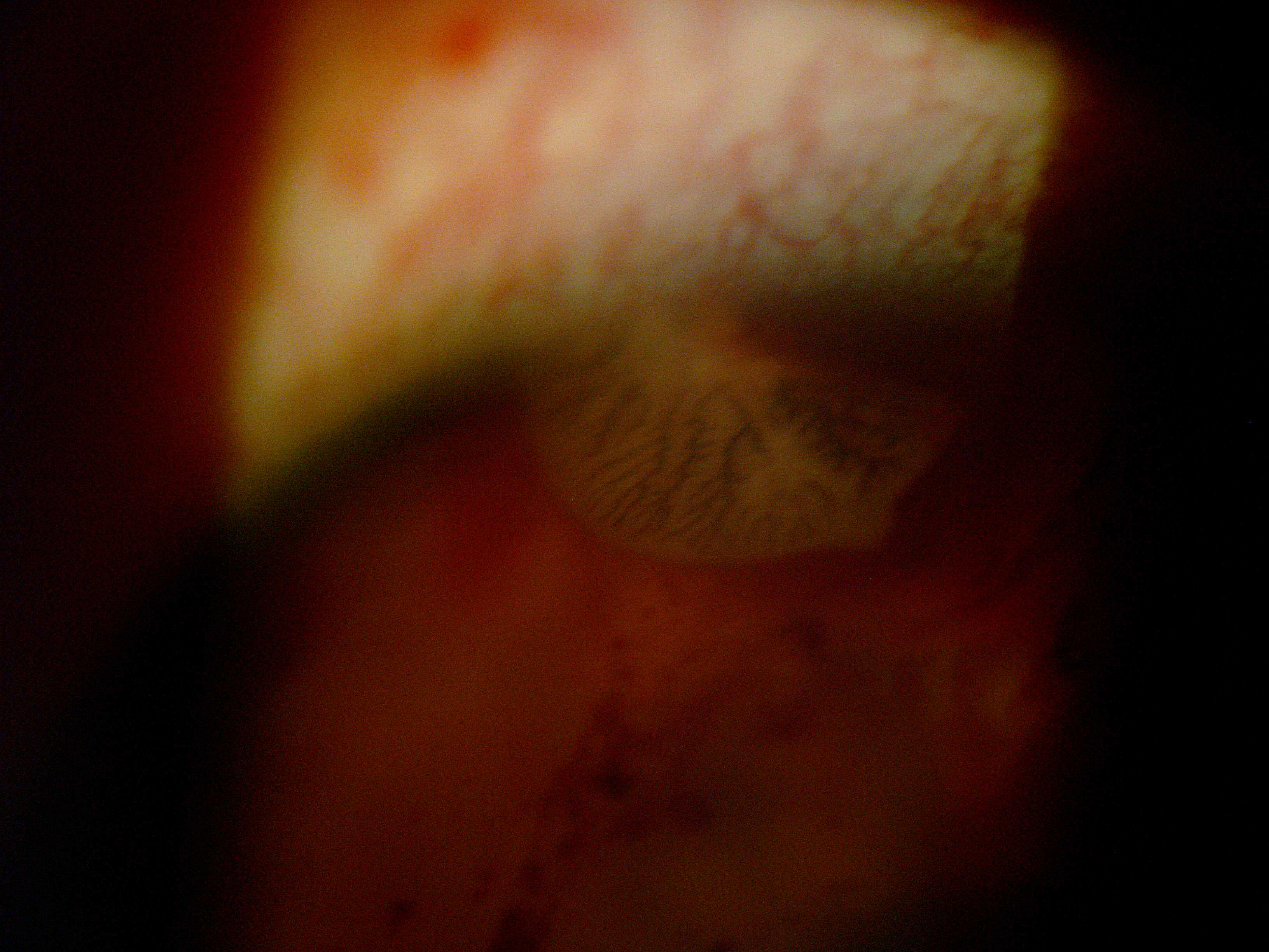
The patient presented to Dr. Pablo Guzmán-Salas, at the Institute of Ophthalmology in Mexico City, after experiencing 3 weeks of decreased visual acuity and pain in his right eye. Upon slit-lamp examination, Dr. Guzman-Salas observed a parasitic fluke (trematode) moving freely in the anterior chamber. A video of the mobile creature accompanied a case report that was published on September 21 in The New England Journal of Medicine.
Extensive ocular damage and inflammation from the worm were also noted, including corneal edema, blood in the anterior chamber, high IOP, multiple iris perforations, and zones of retinal ischemia on the posterior segment.
The report details that the trematode was migrating from the anterior to posterior chambers through the iris perforations it had created.
 Close-up of the live parasite inside the patient's eye. Picture courtesy of Dr. Pablo José Guzmán-Salas
The teen was treated with oral praziquantel and underwent a lensectomy, vitrectomy, and surgical removal of the worm, which had to be cut into "multiple pieces" for extraction. A more specific identification of the type of tremotode—a class that includes an estimated 24,000 species—was not determined.
At 6 months postop, the patient’s vision in the affected eye remained at hand motions.
Curiously, the treating physicians remain unsure how the boy became infected with the parasite. According to his self-reported history, he did not eat any foods that could have put him at risk, and he had not been swimming in any bodies of water. There were no other worms, cysts or eggs found elsewhere in his body.